|
|
|
|
 Description
|
|
Compounds
|
||||||||||||||||||||||||||||||||||||
Chains, Units
Summary Information (see also Sequences/Alignments below) |
Ligands, Modified Residues, Ions (3, 8)
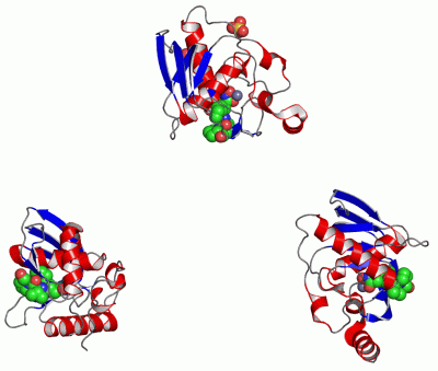
Asymmetric Unit (3, 8)
|
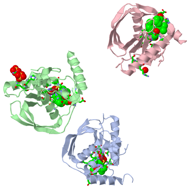
Sites (8, 8)
Asymmetric Unit (8, 8)
|
SS Bonds (0, 0)| (no "SS Bond" information available for 1LRU) |
Cis Peptide Bonds (3, 3)
Asymmetric Unit
|
||||||||||||||||
SAPs(SNPs)/Variants (0, 0)| (no "SAP(SNP)/Variant" information available for 1LRU) |
PROSITE Motifs (0, 0)| (no "PROSITE Motif" information available for 1LRU) |
Exons (0, 0)| (no "Exon" information available for 1LRU) |
Sequences/Alignments
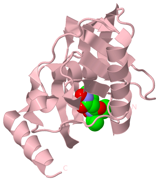
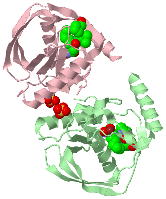
Asymmetric UnitChain A from PDB Type:PROTEIN Length:164 aligned with DEF_ECOLI | P0A6K3 from UniProtKB/Swiss-Prot Length:169 Alignment length:164 11 21 31 41 51 61 71 81 91 101 111 121 131 141 151 161 DEF_ECOLI 2 SVLQVLHIPDERLRKVAKPVEEVNAEIQRIVDDMFETMYAEEGIGLAATQVDIHQRIIVIDVSENRDERLVLINPELLEKSGETGIEEGCLSIPEQRALVPRAEKVKIRALDRDGKPFELEADGLLAICIQHEMDHLVGKLFMDYLSPLKQQRIRQKVEKLDRL 165 SCOP domains d1lrua_ A: Peptide deformylase SCOP domains CATH domains 1lruA00 A:1-164 Peptide Deformylase CATH domains Pfam domains -------------------------------------------------------------------------------------------------------------------------------------------------------------------- Pfam domains SAPs(SNPs) -------------------------------------------------------------------------------------------------------------------------------------------------------------------- SAPs(SNPs) PROSITE -------------------------------------------------------------------------------------------------------------------------------------------------------------------- PROSITE Transcript -------------------------------------------------------------------------------------------------------------------------------------------------------------------- Transcript 1lru A 1 SVLQVLHIPDERLRKVAKPVEEVNAEIQRIVDDMFETMYAEEGIGLAATQVDIHQRIIVIDVSENRDERLVLINPELLEKSGETGIEEGCLSIPEQRALVPRAEKVKIRALDRDGKPFELEADGLLAICIQHEMDHLVGKLFMDYLSPLKQQRIRQKVEKLDRL 164 10 20 30 40 50 60 70 80 90 100 110 120 130 140 150 160 Chain B from PDB Type:PROTEIN Length:164 aligned with DEF_ECOLI | P0A6K3 from UniProtKB/Swiss-Prot Length:169 Alignment length:164 11 21 31 41 51 61 71 81 91 101 111 121 131 141 151 161 DEF_ECOLI 2 SVLQVLHIPDERLRKVAKPVEEVNAEIQRIVDDMFETMYAEEGIGLAATQVDIHQRIIVIDVSENRDERLVLINPELLEKSGETGIEEGCLSIPEQRALVPRAEKVKIRALDRDGKPFELEADGLLAICIQHEMDHLVGKLFMDYLSPLKQQRIRQKVEKLDRL 165 SCOP domains d1lrub_ B: Peptide deformylase SCOP domains CATH domains 1lruB00 B:1-164 Peptide Deformylase CATH domains Pfam domains -------------------------------------------------------------------------------------------------------------------------------------------------------------------- Pfam domains SAPs(SNPs) -------------------------------------------------------------------------------------------------------------------------------------------------------------------- SAPs(SNPs) PROSITE -------------------------------------------------------------------------------------------------------------------------------------------------------------------- PROSITE Transcript -------------------------------------------------------------------------------------------------------------------------------------------------------------------- Transcript 1lru B 1 SVLQVLHIPDERLRKVAKPVEEVNAEIQRIVDDMFETMYAEEGIGLAATQVDIHQRIIVIDVSENRDERLVLINPELLEKSGETGIEEGCLSIPEQRALVPRAEKVKIRALDRDGKPFELEADGLLAICIQHEMDHLVGKLFMDYLSPLKQQRIRQKVEKLDRL 164 10 20 30 40 50 60 70 80 90 100 110 120 130 140 150 160 Chain C from PDB Type:PROTEIN Length:161 aligned with DEF_ECOLI | P0A6K3 from UniProtKB/Swiss-Prot Length:169 Alignment length:161 11 21 31 41 51 61 71 81 91 101 111 121 131 141 151 161 DEF_ECOLI 2 SVLQVLHIPDERLRKVAKPVEEVNAEIQRIVDDMFETMYAEEGIGLAATQVDIHQRIIVIDVSENRDERLVLINPELLEKSGETGIEEGCLSIPEQRALVPRAEKVKIRALDRDGKPFELEADGLLAICIQHEMDHLVGKLFMDYLSPLKQQRIRQKVEKL 162 SCOP domains d1lruc_ C: Peptide deformylase SCOP domains CATH domains 1lruC00 C:1-161 Peptide Deformylase CATH domains Pfam domains (1) -Pep_deformylase-1lruC01 C:2-152 --------- Pfam domains (1) Pfam domains (2) -Pep_deformylase-1lruC02 C:2-152 --------- Pfam domains (2) Pfam domains (3) -Pep_deformylase-1lruC03 C:2-152 --------- Pfam domains (3) SAPs(SNPs) ----------------------------------------------------------------------------------------------------------------------------------------------------------------- SAPs(SNPs) PROSITE ----------------------------------------------------------------------------------------------------------------------------------------------------------------- PROSITE Transcript ----------------------------------------------------------------------------------------------------------------------------------------------------------------- Transcript 1lru C 1 SVLQVLHIPDERLRKVAKPVEEVNAEIQRIVDDMFETMYAEEGIGLAATQVDIHQRIIVIDVSENRDERLVLINPELLEKSGETGIEEGCLSIPEQRALVPRAEKVKIRALDRDGKPFELEADGLLAICIQHEMDHLVGKLFMDYLSPLKQQRIRQKVEKL 161 10 20 30 40 50 60 70 80 90 100 110 120 130 140 150 160
|
||||||||||||||||||||
SCOP Domains (1, 3)
Asymmetric Unit
 |
CATH Domains (1, 3)
Asymmetric Unit
|
Pfam Domains (1, 3)
Asymmetric Unit
|
Gene Ontology (12, 12)|
Asymmetric Unit(hide GO term definitions) Chain A,B,C (DEF_ECOLI | P0A6K3)
|
||||||||||||||||||||||||||||||||||||||||||||||||||||||||||||||||||||||||||||||||||||||||||
Interactive Views
|
|||||||||||||||||||||||||||||||||||||||||||||||||||||||||||||||||||||||||||||||||||||||||||||||||||||||||||||||||||||||||||||||||||||||||||||||||||||||||||||||||||||||||||||||||||||||||||||||||||||||||||||||||||||||||||||||||||||
Still Images
|
||||||||||||||||
Databases
|
||||||||||||||||||||||||||||||||||||||||||||||||||||||||||||||||||||||||||||||||||||||||||||||||||||||||||||||||||||||||||||||||||||||||||||||||||||||||||||||||
Analysis Tools
|
|||||||||||||||||||||||||||||||||||||||||||||||||||||||||||||
Entries Sharing at Least One Protein Chain (UniProt ID)
Related Entries Specified in the PDB File
|
|