Show PDB file:   
         Plain Text   HTML   (compressed file size)
QuickSearch:   
by PDB,NDB,UniProt,PROSITE Code or Search Term(s)  
(-)NMR Structure - model 1
(-)NMR Structure - model 1, sites
(-)NMR Structure - all models
collapse expand < >
Image NMR Structure - model 1
NMR Structure - model 1  (Jmol Viewer)
Image NMR Structure - model 1, sites
NMR Structure - model 1, sites  (Jmol Viewer)
Image NMR Structure - all models
NMR Structure - all models  (Jmol Viewer)

(-) Description

Title :  SOLUTION STRUCTURE DETERMINATION OF THE FULLY OXIDIZED DOUBLE MUTANT K9-10A CYTOCHROME C7 FROM DESULFUROMONAS ACETOXIDANS, ENSEMBLE OF 35 STRUCTURES
 
Authors :  M. Assfalg, I. Bertini, P. Turano, M. Bruschi, M. C. Durand, M. T. Giudici-Orticoni, A. Dolla
Date :  28 Feb 02  (Deposition) - 13 Mar 02  (Release) - 24 Feb 09  (Revision)
Method :  SOLUTION NMR
Resolution :  NOT APPLICABLE
Chains :  NMR Structure  :  A  (35x)
Keywords :  Automatic Assignment, Cytochrome C7, Electron Transfer, Multiheme Cytochromes, Nmr Solution Structures, Oxygen Storage/Transport Complex (Keyword Search: [Gene Ontology, PubMed, Web (Google))
 
Reference :  M. Assfalg, I. Bertini, P. Turano, M. Bruschi, M. C. Durand, M. T. Giudici-Orticoni, A. Dolla
A Quick Solution Structure Determination Of The Fully Oxidized Double Mutant K9-10A Cytochrome C7 From Desulfuromonas Acetoxidans And Mechanistic Implications.
J. Biomol. Nmr V. 22 107 2002
PubMed-ID: 11883773  |  Reference-DOI: 10.1023/A:1014202405862
(for further references see the PDB file header)

(-) Compounds

Molecule 1 - CYTOCHROME C7
    ChainsA
    EngineeredYES
    Expression SystemDESULFOVIBRIO DESULFURICANS
    Expression System StrainG201
    Expression System Taxid876
    MutationYES
    Organism ScientificDESULFUROMONAS ACETOXIDANS
    Organism Taxid891
    SynonymCYTOCHROME C3, CYTOCHROME C551.5

 Structural Features

(-) Chains, Units

  
NMR Structure (35x)

Summary Information (see also Sequences/Alignments below)

(-) Ligands, Modified Residues, Ions  (1, 3)

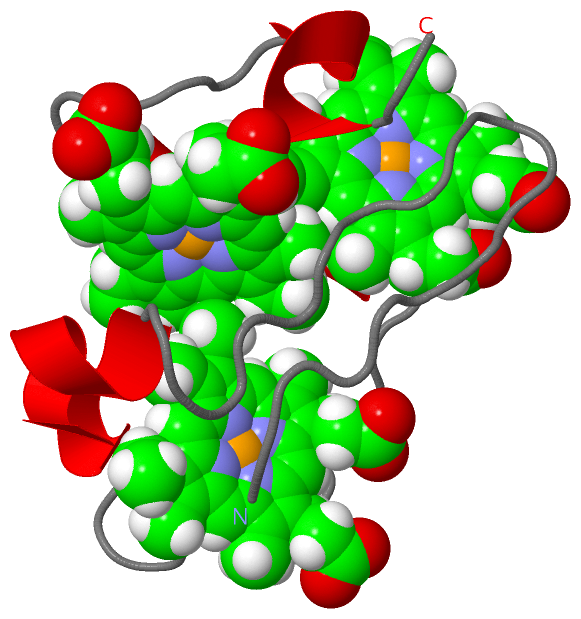
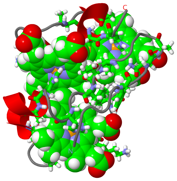
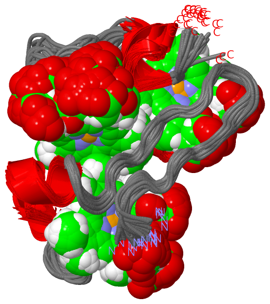
NMR Structure (1, 3)
No.NameCountTypeFull Name
1HEC3Ligand/IonHEME C

(-) Sites  (3, 3)

NMR Structure (3, 3)
No.NameEvidenceResiduesDescription
1AC1SOFTWARETYR A:6 , HIS A:17 , ALA A:21 , CYS A:26 , CYS A:29 , HIS A:30 , PRO A:34 , ALA A:35 , LYS A:36 , ILE A:37 , ILE A:39 , HEC A:153BINDING SITE FOR RESIDUE HEC A 130
2AC2SOFTWAREVAL A:13 , THR A:14 , PHE A:15 , HIS A:20 , CYS A:29 , CYS A:49 , CYS A:52 , HIS A:53 , ASN A:56 , HEC A:130BINDING SITE FOR RESIDUE HEC A 153
3AC3SOFTWAREASN A:8 , ALA A:9 , VAL A:13 , ASP A:40 , LYS A:41 , ALA A:44 , HIS A:45 , THR A:60 , CYS A:62 , CYS A:65 , HIS A:66BINDING SITE FOR RESIDUE HEC A 166

(-) SS Bonds  (0, 0)

(no "SS Bond" information available for 1L3O)

(-) Cis Peptide Bonds  (0, 0)

(no "Cis Peptide Bond" information available for 1L3O)

 Sequence-Structure Mapping

(-) SAPs(SNPs)/Variants  (0, 0)

(no "SAP(SNP)/Variant" information available for 1L3O)

(-) PROSITE Motifs  (0, 0)

(no "PROSITE Motif" information available for 1L3O)

(-) Exons   (0, 0)

(no "Exon" information available for 1L3O)

(-) Sequences/Alignments

NMR Structure
   Reformat: Number of residues per line =  ('0' or empty: single-line sequence representation)
  Number of residues per labelling interval =   
  UniProt sequence: complete  aligned part    
   Show mapping: SCOP domains CATH domains Pfam domains Secondary structure (by author)
SAPs(SNPs) PROSITE motifs Exons
(details for a mapped element are shown in a popup box when the mouse pointer rests over it)
Chain A from PDB  Type:PROTEIN  Length:68
 aligned with CYC3_DESAC | P00137 from UniProtKB/Swiss-Prot  Length:68

    Alignment length:68
                                    10        20        30        40        50        60        
            CYC3_DESAC    1 ADVVTYENKKGNVTFDHKAHAEKLGCDACHEGTPAKIAIDKKSAHKDACKTCHKSNNGPTKCGGCHIK 68
               SCOP domains d1l3oa_ A: Cytochrome c7 (cytochrome c551.5, PpcA)                   SCOP domains
               CATH domains 1l3oA00 A:1-68 Cytochrome C3                                         CATH domains
               Pfam domains Cytochrom_CIII-1l3oA01 A:1-68                                        Pfam domains
         Sec.struct. author ...................hhhhhhhhhhh...........hhhhhh.hhhhhhh.....hhhhh... Sec.struct. author
                 SAPs(SNPs) -------------------------------------------------------------------- SAPs(SNPs)
                    PROSITE -------------------------------------------------------------------- PROSITE
                 Transcript -------------------------------------------------------------------- Transcript
                  1l3o A  1 ADVVTYENAAGNVTFDHKAHAEKLGCDACHEGTPAKIAIDKKSAHKDACKTCHKSNNGPTKCGGCHIK 68
                                    10        20        30        40        50        60        

   Legend:   → Mismatch (orange background)
  - → Gap (green background, '-', border residues have a numbering label)
    → Modified Residue (blue background, lower-case, 'x' indicates undefined single-letter code, labelled with number + name)
  x → Chemical Group (purple background, 'x', labelled with number + name, e.g. ACE or NH2)
  extra numbering lines below/above indicate numbering irregularities and modified residue names etc., number ends below/above '|'

 Classification and Annotation

(-) SCOP Domains  (1, 1)

NMR Structure

(-) CATH Domains  (1, 1)

NMR Structure
(-)
Class: Alpha Beta (26913)

(-) Pfam Domains  (1, 1)

NMR Structure

(-) Gene Ontology  (5, 5)

NMR Structure(hide GO term definitions)
Chain A   (CYC3_DESAC | P00137)
molecular function
    GO:0009055    electron carrier activity    Any molecular entity that serves as an electron acceptor and electron donor in an electron transport chain. An electron transport chain is a process in which a series of electron carriers operate together to transfer electrons from donors to any of several different terminal electron acceptors to generate a transmembrane electrochemical gradient.
    GO:0020037    heme binding    Interacting selectively and non-covalently with heme, any compound of iron complexed in a porphyrin (tetrapyrrole) ring.
    GO:0046872    metal ion binding    Interacting selectively and non-covalently with any metal ion.
biological process
    GO:0009061    anaerobic respiration    The enzymatic release of energy from inorganic and organic compounds (especially carbohydrates and fats) which uses compounds other than oxygen (e.g. nitrate, sulfate) as the terminal electron acceptor.
    GO:0055114    oxidation-reduction process    A metabolic process that results in the removal or addition of one or more electrons to or from a substance, with or without the concomitant removal or addition of a proton or protons.

 Visualization

(-) Interactive Views

NMR Structure
  Complete Structure
    Jena3D(integrated viewing of ligand, site, SAP, PROSITE, SCOP information)
    WebMol | AstexViewer[tm]@PDBe
(Java Applets, require no local installation except for Java; loading may be slow)
    STRAP
(Java WebStart application, automatic local installation, requires Java; full application with system access!)
    RasMol
(require local installation)
    Molscript (VRML)
(requires installation of a VRML viewer; select preferred view via VRML and generate a mono or stereo PDF format file)
 
  Ligands, Modified Residues, Ions
    HEC  [ RasMol | Jena3D ]  +environment [ RasMol | Jena3D ]
 
  Sites
    AC1  [ RasMol ]  +environment [ RasMol ]
    AC2  [ RasMol ]  +environment [ RasMol ]
    AC3  [ RasMol ]  +environment [ RasMol ]
 
  Cis Peptide Bonds
(no "Cis Peptide Bonds" information available for 1l3o)
 

(-) Still Images

Jmol
  protein: cartoon or spacefill or dots and stick; nucleic acid: cartoon and stick; ligands: spacefill; active site: stick
Molscript
  protein, nucleic acid: cartoon; ligands: spacefill; active site: ball and stick

 Databases and Analysis Tools

(-) Databases

Access by PDB/NDB ID
  1l3o
    Family and Domain InformationProDom | SYSTERS
    General Structural InformationGlycoscienceDB | MMDB | NDB | OCA | PDB | PDBe | PDBj | PDBsum | PDBWiki | PQS | PROTEOPEDIA
    Orientation in MembranesOPM
    Protein SurfaceSURFACE
    Secondary StructureDSSP (structure derived) | HSSP (homology derived)
    Structural GenomicsGeneCensus
    Structural NeighboursCE | VAST
    Structure ClassificationCATH | Dali | SCOP
    Validation and Original DataBMRB Data View | BMRB Restraints Grid | EDS | PROCHECK | RECOORD | WHAT_CHECK
 
Access by UniProt ID/Accession number
  CYC3_DESAC | P00137
    Comparative Protein Structure ModelsModBase
    Genomic InformationEnsembl
    Protein-protein InteractionDIP
    Sequence, Family and Domain InformationInterPro | Pfam | SMART | UniProtKB/SwissProt
 
Access by Enzyme Classificator   (EC Number)
  (no 'Enzyme Classificator' available)
    General Enzyme InformationBRENDA | EC-PDB | Enzyme | IntEnz
    PathwayKEGG | MetaCyc
 
Access by Disease Identifier   (MIM ID)
  (no 'MIM ID' available)
    Disease InformationOMIM
 
Access by GenAge ID
  (no 'GenAge ID' available)
    Age Related InformationGenAge

(-) Analysis Tools

Access by PDB/NDB ID
    Domain InformationXDom
    Interatomic Contacts of Structural UnitsCSU
    Ligand-protein ContactsLPC
    Protein CavitiescastP
    Sequence and Secondary StructurePDBCartoon
    Structure AlignmentSTRAP(Java WebStart application, automatic local installation, requires Java; full application with system access!)
    Structure and Sequence BrowserSTING
 
Access by UniProt ID/Accession number
  CYC3_DESAC | P00137
    Protein Disorder PredictionDisEMBL | FoldIndex | GLOBPLOT (for more information see DisProt)

 Related Entries

(-) Entries Sharing at Least One Protein Chain (UniProt ID)

UniProtKB/Swiss-Prot
        CYC3_DESAC | P001371ehj 1f22 1hh5 1kwj 1lm2 1new

(-) Related Entries Specified in the PDB File

5279 CONTAINS THE PROTON SHIFTS FOR THE PRESENT STRUCTURE
1ehj CONTAINS 35 NMR STRUCTURES OF THE FULLY REDUCED NATIVE PROTEIN
1hh5 CONTAINS THE X-RAY STRUCTURE OF THE NATIVE PROTEIN
1kwj CONTAINS THE ENERGY MINIMIZED AVERAGE NMR STRUCTURE FROM THE PRESENT FAMILY OF STRUCTURES
1new CONTAINS 18 NMR STRUCTURES OF THE FULLY OXIDIZED NATIVE CYTOCHROME C7
2new CONTAINS 17 NMR STRUCTURES OF THE FULLY OXIDIZED NATIVE CYTOCHROME C7