|
|
|
|
 Description
|
|
Compounds
|
||||||||||||||||||||||||||||||||||||||||||||||||||||||||||||||||
Chains, Units
Summary Information (see also Sequences/Alignments below) |
Ligands, Modified Residues, Ions (2, 15)| Asymmetric/Biological Unit (2, 15) |
Sites (1, 1)
Asymmetric Unit (1, 1)
|
SS Bonds (0, 0)| (no "SS Bond" information available for 1JAD) |
Cis Peptide Bonds (0, 0)| (no "Cis Peptide Bond" information available for 1JAD) |
SAPs(SNPs)/Variants (0, 0)| (no "SAP(SNP)/Variant" information available for 1JAD) |
PROSITE Motifs (0, 0)| (no "PROSITE Motif" information available for 1JAD) |
Exons (0, 0)| (no "Exon" information available for 1JAD) |
Sequences/Alignments
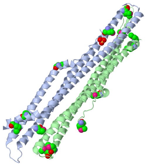
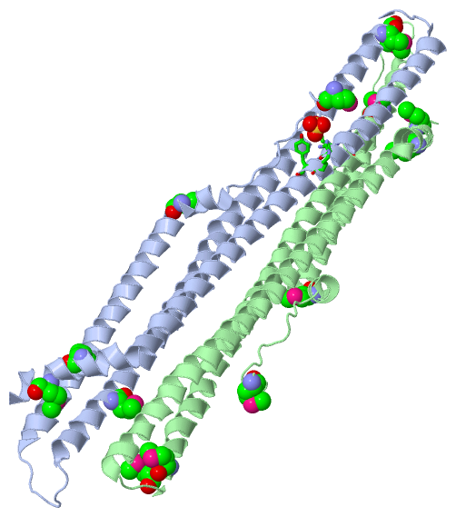
Asymmetric/Biological UnitChain A from PDB Type:PROTEIN Length:242 aligned with Q91086_MELGA | Q91086 from UniProtKB/TrEMBL Length:1211 Alignment length:275 889 899 909 919 929 939 949 959 969 979 989 999 1009 1019 1029 1039 1049 1059 1069 1079 1089 1099 1109 1119 1129 1139 1149 Q91086_MELGA 880 NMKEVTQLPEPQTASLAELQQMKLFLKLLKKQEKELKELERKGSKRREELLQKYSVLFLEPVYPRGKKRSMHSRKTQKKRSLTTGDVGTCMQPVEMAEKLDSQVVELKERLEMELIHLGEEYHDGIRRRKEQHATEQTAKITELAREKQIAELKALKESSESNIKDIKKKLEAKRLDRIQVMMRSTSDKAAQERLKKEINNSHIQEVVQTIKLLTEKTARYQQKLEEKQAENLRAIQEKEGQLQQEAVAEYEEKLKTLTVEVQEMVKNYMKEVFP 1154 SCOP domains d1jada_ A: C-terminal domain of PLC-beta SCOP domains CATH domains 1jadA00 A:3-244 [code=1.20.1230.10, no name defined] CATH domains Pfam domains ----------------------------------------------------------------------------------------------------------------------------------------------------------------------------------------------------------------------------------------------------------------------------------- Pfam domains SAPs(SNPs) ----------------------------------------------------------------------------------------------------------------------------------------------------------------------------------------------------------------------------------------------------------------------------------- SAPs(SNPs) PROSITE ----------------------------------------------------------------------------------------------------------------------------------------------------------------------------------------------------------------------------------------------------------------------------------- PROSITE Transcript ----------------------------------------------------------------------------------------------------------------------------------------------------------------------------------------------------------------------------------------------------------------------------------- Transcript 1jad A 3 NmKEVTQLPEPQTASLAELQQmKLFLKLLKKQEKELKELERKGSKRREELLQKYSVLFLEPVYPRG---------------------------------LDSQVVELKERLEmELIHLGEEYHDGIRRRKEQHATEQTAKITELAREKQIAELKALKESSESNIKDIKKKLEAKRLDRIQVmmRSTSDKAAQERLKKEINNSHIQEVVQTIKLLTEKTARYQQKLEEKQAENLRAIQEKEGQLQQEAVAEYEEKLKTLTVEVQEmVKNYmKEVFP 244 | 12 22 | 32 42 52 62 | - - - 69 79 | 89 99 109 119 129 139 149 || 159 169 179 189 199 209 219 229 | 239 | 24-MSE 68 69 82-MSE 151-MSE 234-MSE| 4-MSE 152-MSE 239-MSE Chain B from PDB Type:PROTEIN Length:242 aligned with Q91086_MELGA | Q91086 from UniProtKB/TrEMBL Length:1211 Alignment length:275 889 899 909 919 929 939 949 959 969 979 989 999 1009 1019 1029 1039 1049 1059 1069 1079 1089 1099 1109 1119 1129 1139 1149 Q91086_MELGA 880 NMKEVTQLPEPQTASLAELQQMKLFLKLLKKQEKELKELERKGSKRREELLQKYSVLFLEPVYPRGKKRSMHSRKTQKKRSLTTGDVGTCMQPVEMAEKLDSQVVELKERLEMELIHLGEEYHDGIRRRKEQHATEQTAKITELAREKQIAELKALKESSESNIKDIKKKLEAKRLDRIQVMMRSTSDKAAQERLKKEINNSHIQEVVQTIKLLTEKTARYQQKLEEKQAENLRAIQEKEGQLQQEAVAEYEEKLKTLTVEVQEMVKNYMKEVFP 1154 SCOP domains d1jadb_ B: C-terminal domain of PLC-beta SCOP domains CATH domains 1jadB00 B:284-525 [code=1.20.1230.10, no name defined] CATH domains Pfam domains ----------------------------------------------------------------------------------------------------------------------------------------------------------------------------------------------------------------------------------------------------------------------------------- Pfam domains SAPs(SNPs) ----------------------------------------------------------------------------------------------------------------------------------------------------------------------------------------------------------------------------------------------------------------------------------- SAPs(SNPs) PROSITE ----------------------------------------------------------------------------------------------------------------------------------------------------------------------------------------------------------------------------------------------------------------------------------- PROSITE Transcript ----------------------------------------------------------------------------------------------------------------------------------------------------------------------------------------------------------------------------------------------------------------------------------- Transcript 1jad B 284 NmKEVTQLPEPQTASLAELQQmKLFLKLLKKQEKELKELERKGSKRREELLQKYSVLFLEPVYPRG---------------------------------LDSQVVELKERLEmELIHLGEEYHDGIRRRKEQHATEQTAKITELAREKQIAELKALKESSESNIKDIKKKLEAKRLDRIQVmmRSTSDKAAQERLKKEINNSHIQEVVQTIKLLTEKTARYQQKLEEKQAENLRAIQEKEGQLQQEAVAEYEEKLKTLTVEVQEmVKNYmKEVFP 525 | 293 303 | 313 323 333 343 | - - - 350 360 | 370 380 390 400 410 420 430 || 440 450 460 470 480 490 500 510 | 520 | 305-MSE 349 350 363-MSE 432-MSE 515-MSE| 285-MSE 433-MSE 520-MSE
|
||||||||||||||||||||
SCOP Domains (1, 2)
Asymmetric/Biological Unit
 |
CATH Domains (1, 2)
Asymmetric/Biological Unit
|
Pfam Domains (0, 0)| (no "Pfam Domain" information available for 1JAD) |
Gene Ontology (11, 11)|
Asymmetric/Biological Unit(hide GO term definitions) Chain A,B (Q91086_MELGA | Q91086)
|
||||||||||||||||||||||||||||||||||||||||||||||||||||||||||||||||||||||||||||||||||||
Interactive Views
|
|||||||||||||||||||||||||||||||||||||||||||||||||||||||||||||||||||||||||||||||||||||||||||||||||||||||||||||||||||||||||||||
Still Images
|
||||||||||||||||
Databases
|
||||||||||||||||||||||||||||||||||||||||||||||||||||||||||||||||||||||||||||||||||||||||||||||||||||||||||||||||||||||||||||||||||||||||||||||||||||||||||||||||
Analysis Tools
|
|||||||||||||||||||||||||||||||||||||||||||||||||||||||||||||
Entries Sharing at Least One Protein Chain (UniProt ID)
Related Entries Specified in the PDB File
|
|