|
|
|
|
 Description
|
|
Compounds
|
||||||||||||||||||||||||||||||||||||||||||||||||
Chains, Units
Summary Information (see also Sequences/Alignments below) |
Ligands, Modified Residues, Ions (1, 4)
Asymmetric Unit (1, 4)
|
Sites (4, 4)
Asymmetric Unit (4, 4)
|
SS Bonds (0, 0)| (no "SS Bond" information available for 1G28) |
Cis Peptide Bonds (0, 0)| (no "Cis Peptide Bond" information available for 1G28) |
SAPs(SNPs)/Variants (0, 0)| (no "SAP(SNP)/Variant" information available for 1G28) |
PROSITE Motifs (0, 0)| (no "PROSITE Motif" information available for 1G28) |
Exons (0, 0)| (no "Exon" information available for 1G28) |
Sequences/Alignments
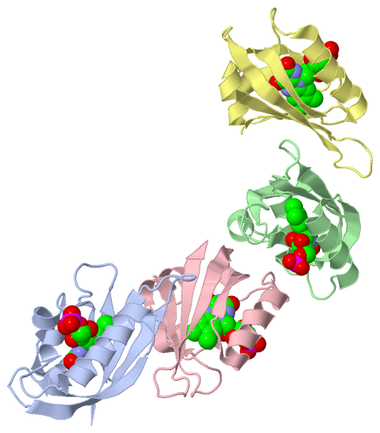
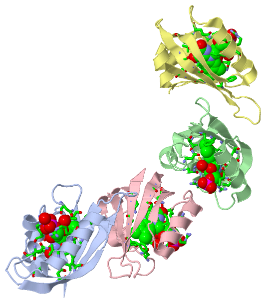
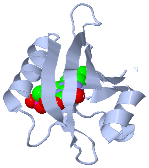
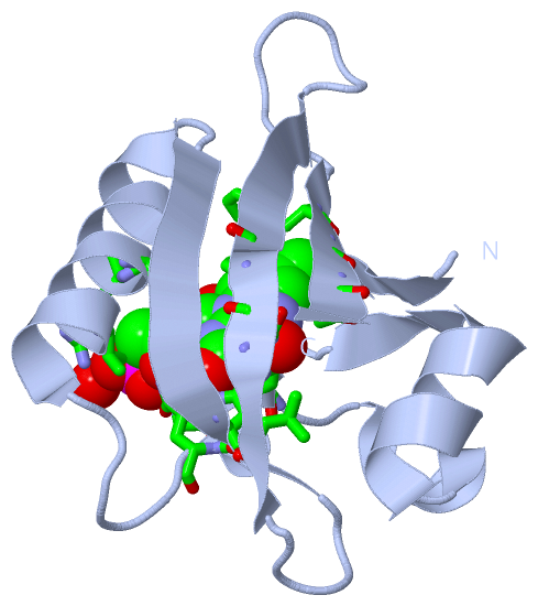
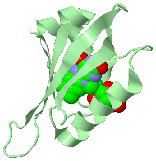
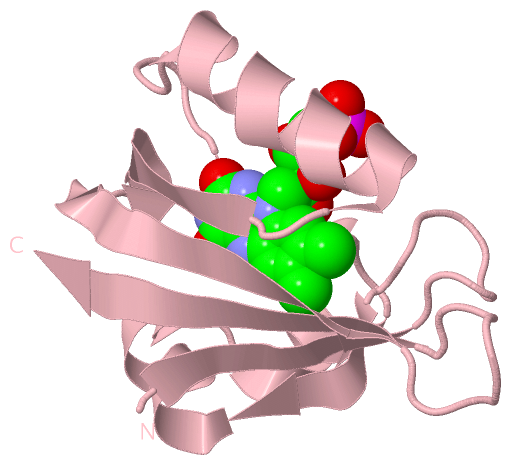
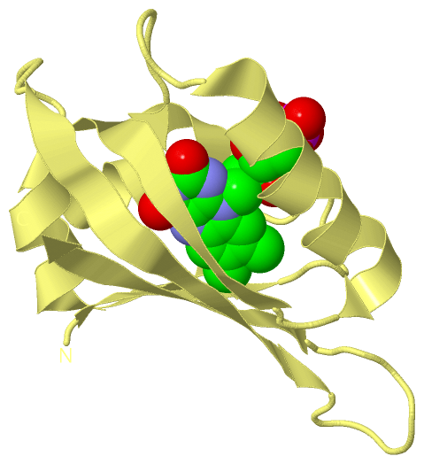
Asymmetric UnitChain A from PDB Type:PROTEIN Length:104 aligned with Q9ZWQ6_ADICA | Q9ZWQ6 from UniProtKB/TrEMBL Length:1465 Alignment length:104 938 948 958 968 978 988 998 1008 1018 1028 Q9ZWQ6_ADICA 929 KSFVITDPRLPDNPIIFASDRFLELTEYTREEVLGNNCRFLQGRGTDRKAVQLIRDAVKEQRDVTVQVLNYTKGGRAFWNLFHLQVMRDENGDVQYFIGVQQEM 1032 SCOP domains d1g28a_ A: Photoreceptor phy3 flavin-binding domain, lov2 SCOP domains CATH domains 1g28A00 A:929-1032 [code=3.30.450.20, no name defined] CATH domains Pfam domains -------------------------------------------------------------------------------------------------------- Pfam domains SAPs(SNPs) -------------------------------------------------------------------------------------------------------- SAPs(SNPs) PROSITE -------------------------------------------------------------------------------------------------------- PROSITE Transcript -------------------------------------------------------------------------------------------------------- Transcript 1g28 A 929 KSFVITDPRLPDNPIIFASDRFLELTEYTREEVLGNNCRFLQGRGTDRKAVQLIRDAVKEQRDVTVQVLNYTKGGRAFWNLFHLQVMRDENGDVQYFIGVQQEM 1032 938 948 958 968 978 988 998 1008 1018 1028 Chain B from PDB Type:PROTEIN Length:104 aligned with Q9ZWQ6_ADICA | Q9ZWQ6 from UniProtKB/TrEMBL Length:1465 Alignment length:104 938 948 958 968 978 988 998 1008 1018 1028 Q9ZWQ6_ADICA 929 KSFVITDPRLPDNPIIFASDRFLELTEYTREEVLGNNCRFLQGRGTDRKAVQLIRDAVKEQRDVTVQVLNYTKGGRAFWNLFHLQVMRDENGDVQYFIGVQQEM 1032 SCOP domains d1g28b_ B: Photoreceptor phy3 flavin-binding domain, lov2 SCOP domains CATH domains 1g28B00 B:929-1032 [code=3.30.450.20, no name defined] CATH domains Pfam domains -------------------------------------------------------------------------------------------------------- Pfam domains SAPs(SNPs) -------------------------------------------------------------------------------------------------------- SAPs(SNPs) PROSITE -------------------------------------------------------------------------------------------------------- PROSITE Transcript -------------------------------------------------------------------------------------------------------- Transcript 1g28 B 929 KSFVITDPRLPDNPIIFASDRFLELTEYTREEVLGNNCRFLQGRGTDRKAVQLIRDAVKEQRDVTVQVLNYTKGGRAFWNLFHLQVMRDENGDVQYFIGVQQEM 1032 938 948 958 968 978 988 998 1008 1018 1028 Chain C from PDB Type:PROTEIN Length:104 aligned with Q9ZWQ6_ADICA | Q9ZWQ6 from UniProtKB/TrEMBL Length:1465 Alignment length:104 938 948 958 968 978 988 998 1008 1018 1028 Q9ZWQ6_ADICA 929 KSFVITDPRLPDNPIIFASDRFLELTEYTREEVLGNNCRFLQGRGTDRKAVQLIRDAVKEQRDVTVQVLNYTKGGRAFWNLFHLQVMRDENGDVQYFIGVQQEM 1032 SCOP domains d1g28c_ C: Photoreceptor phy3 flavin-binding domain, lov2 SCOP domains CATH domains 1g28C00 C:929-1032 [code=3.30.450.20, no name defined] CATH domains Pfam domains -------------------------------------------------------------------------------------------------------- Pfam domains SAPs(SNPs) -------------------------------------------------------------------------------------------------------- SAPs(SNPs) PROSITE -------------------------------------------------------------------------------------------------------- PROSITE Transcript -------------------------------------------------------------------------------------------------------- Transcript 1g28 C 929 KSFVITDPRLPDNPIIFASDRFLELTEYTREEVLGNNCRFLQGRGTDRKAVQLIRDAVKEQRDVTVQVLNYTKGGRAFWNLFHLQVMRDENGDVQYFIGVQQEM 1032 938 948 958 968 978 988 998 1008 1018 1028 Chain D from PDB Type:PROTEIN Length:104 aligned with Q9ZWQ6_ADICA | Q9ZWQ6 from UniProtKB/TrEMBL Length:1465 Alignment length:104 938 948 958 968 978 988 998 1008 1018 1028 Q9ZWQ6_ADICA 929 KSFVITDPRLPDNPIIFASDRFLELTEYTREEVLGNNCRFLQGRGTDRKAVQLIRDAVKEQRDVTVQVLNYTKGGRAFWNLFHLQVMRDENGDVQYFIGVQQEM 1032 SCOP domains d1g28d_ D: Photoreceptor phy3 flavin-binding domain, lov2 SCOP domains CATH domains 1g28D00 D:929-1032 [code=3.30.450.20, no name defined] CATH domains Pfam domains -------------------------------------------------------------------------------------------------------- Pfam domains SAPs(SNPs) -------------------------------------------------------------------------------------------------------- SAPs(SNPs) PROSITE -------------------------------------------------------------------------------------------------------- PROSITE Transcript -------------------------------------------------------------------------------------------------------- Transcript 1g28 D 929 KSFVITDPRLPDNPIIFASDRFLELTEYTREEVLGNNCRFLQGRGTDRKAVQLIRDAVKEQRDVTVQVLNYTKGGRAFWNLFHLQVMRDENGDVQYFIGVQQEM 1032 938 948 958 968 978 988 998 1008 1018 1028
|
||||||||||||||||||||
SCOP Domains (1, 4)
Asymmetric Unit
 |
CATH Domains (1, 4)
Asymmetric Unit
|
Pfam Domains (0, 0)| (no "Pfam Domain" information available for 1G28) |
Gene Ontology (12, 12)|
Asymmetric Unit(hide GO term definitions) Chain A,B,C,D (Q9ZWQ6_ADICA | Q9ZWQ6)
|
||||||||||||||||||||||||||||||||||||||||||||||||||||||||||||||||||||||||||||||||||||||||||
Interactive Views
|
||||||||||||||||||||||||||||||||||||||||||||||||||||||||||||||||||||||||||||||||||||||||||||||||||||||||||||||||||||||||||||||||||||||||||||||||||||||||||||||||||||||||||||
Still Images
|
||||||||||||||||
Databases
|
||||||||||||||||||||||||||||||||||||||||||||||||||||||||||||||||||||||||||||||||||||||||||||||||||||||||||||||||||||||||||||||||||||||||||||||||||||||||||||||||
Analysis Tools
|
|||||||||||||||||||||||||||||||||||||||||||||||||||||||||||||
Entries Sharing at Least One Protein Chain (UniProt ID)
Related Entries Specified in the PDB File
|
|