|
|
|
|
 Description
|
|
Compounds
|
||||||||||||||||||||||||||||||||||||||||||||||||||||||||||||||||||||||||
Chains, Units
Summary Information (see also Sequences/Alignments below) |
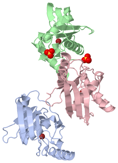
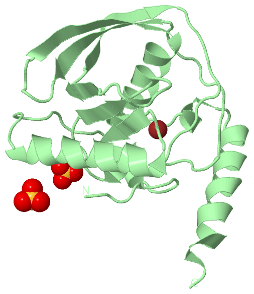
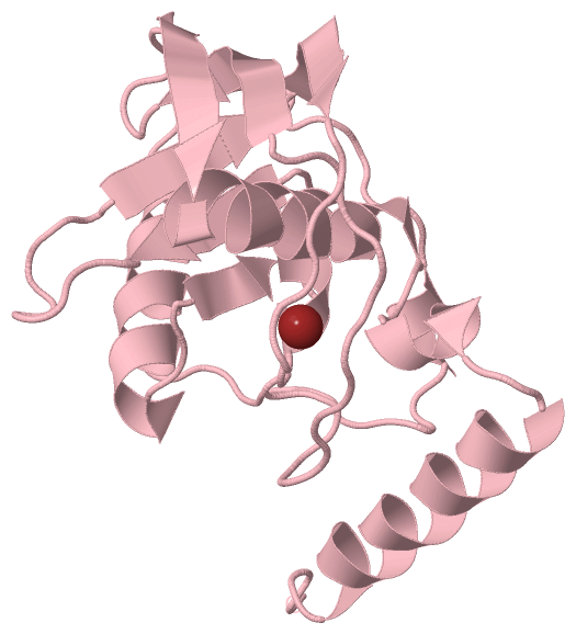
Ligands, Modified Residues, Ions (2, 5)| Asymmetric Unit (2, 5) Biological Unit 1 (0, 0) Biological Unit 2 (1, 2) Biological Unit 3 (0, 0) |
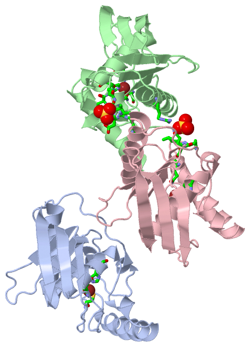
Sites (8, 8)
Asymmetric Unit (8, 8)
|
SS Bonds (0, 0)| (no "SS Bond" information available for 1BS7) |
Cis Peptide Bonds (3, 3)
Asymmetric Unit
|
||||||||||||||||
SAPs(SNPs)/Variants (0, 0)| (no "SAP(SNP)/Variant" information available for 1BS7) |
PROSITE Motifs (0, 0)| (no "PROSITE Motif" information available for 1BS7) |
Exons (0, 0)| (no "Exon" information available for 1BS7) |
Sequences/Alignments
Asymmetric UnitChain A from PDB Type:PROTEIN Length:168 aligned with DEF_ECOLI | P0A6K3 from UniProtKB/Swiss-Prot Length:169 Alignment length:168 11 21 31 41 51 61 71 81 91 101 111 121 131 141 151 161 DEF_ECOLI 2 SVLQVLHIPDERLRKVAKPVEEVNAEIQRIVDDMFETMYAEEGIGLAATQVDIHQRIIVIDVSENRDERLVLINPELLEKSGETGIEEGCLSIPEQRALVPRAEKVKIRALDRDGKPFELEADGLLAICIQHEMDHLVGKLFMDYLSPLKQQRIRQKVEKLDRLKARA 169 SCOP domains d1bs7a_ A: Peptide deformylase SCOP domains CATH domains 1bs7A00 A:1-168 Peptide Deformylase CATH domains Pfam domains ------------------------------------------------------------------------------------------------------------------------------------------------------------------------ Pfam domains SAPs(SNPs) ------------------------------------------------------------------------------------------------------------------------------------------------------------------------ SAPs(SNPs) PROSITE ------------------------------------------------------------------------------------------------------------------------------------------------------------------------ PROSITE Transcript ------------------------------------------------------------------------------------------------------------------------------------------------------------------------ Transcript 1bs7 A 1 SVLQVLHIPDERLRKVAKPVEEVNAEIQRIVDDMFETMYAEEGIGLAATQVDIHQRIIVIDVSENRDERLVLINPELLEKSGETGIEEGCLSIPEQRALVPRAEKVKIRALDRDGKPFELEADGLLAICIQHEMDHLVGKLFMDYLSPLKQQRIRQKVEKLDRLKARA 168 10 20 30 40 50 60 70 80 90 100 110 120 130 140 150 160 Chain B from PDB Type:PROTEIN Length:168 aligned with DEF_ECOLI | P0A6K3 from UniProtKB/Swiss-Prot Length:169 Alignment length:168 11 21 31 41 51 61 71 81 91 101 111 121 131 141 151 161 DEF_ECOLI 2 SVLQVLHIPDERLRKVAKPVEEVNAEIQRIVDDMFETMYAEEGIGLAATQVDIHQRIIVIDVSENRDERLVLINPELLEKSGETGIEEGCLSIPEQRALVPRAEKVKIRALDRDGKPFELEADGLLAICIQHEMDHLVGKLFMDYLSPLKQQRIRQKVEKLDRLKARA 169 SCOP domains d1bs7b_ B: Peptide deformylase SCOP domains CATH domains 1bs7B00 B:501-668 Peptide Deformylase CATH domains Pfam domains ------------------------------------------------------------------------------------------------------------------------------------------------------------------------ Pfam domains SAPs(SNPs) ------------------------------------------------------------------------------------------------------------------------------------------------------------------------ SAPs(SNPs) PROSITE ------------------------------------------------------------------------------------------------------------------------------------------------------------------------ PROSITE Transcript ------------------------------------------------------------------------------------------------------------------------------------------------------------------------ Transcript 1bs7 B 501 SVLQVLHIPDERLRKVAKPVEEVNAEIQRIVDDMFETMYAEEGIGLAATQVDIHQRIIVIDVSENRDERLVLINPELLEKSGETGIEEGCLSIPEQRALVPRAEKVKIRALDRDGKPFELEADGLLAICIQHEMDHLVGKLFMDYLSPLKQQRIRQKVEKLDRLKARA 668 510 520 530 540 550 560 570 580 590 600 610 620 630 640 650 660 Chain C from PDB Type:PROTEIN Length:168 aligned with DEF_ECOLI | P0A6K3 from UniProtKB/Swiss-Prot Length:169 Alignment length:168 11 21 31 41 51 61 71 81 91 101 111 121 131 141 151 161 DEF_ECOLI 2 SVLQVLHIPDERLRKVAKPVEEVNAEIQRIVDDMFETMYAEEGIGLAATQVDIHQRIIVIDVSENRDERLVLINPELLEKSGETGIEEGCLSIPEQRALVPRAEKVKIRALDRDGKPFELEADGLLAICIQHEMDHLVGKLFMDYLSPLKQQRIRQKVEKLDRLKARA 169 SCOP domains d1bs7c_ C: Peptide deformylase SCOP domains CATH domains 1bs7C00 C:1001-1168 Peptide Deformylase CATH domains Pfam domains ------------------------------------------------------------------------------------------------------------------------------------------------------------------------ Pfam domains SAPs(SNPs) ------------------------------------------------------------------------------------------------------------------------------------------------------------------------ SAPs(SNPs) PROSITE ------------------------------------------------------------------------------------------------------------------------------------------------------------------------ PROSITE Transcript ------------------------------------------------------------------------------------------------------------------------------------------------------------------------ Transcript 1bs7 C 1001 SVLQVLHIPDERLRKVAKPVEEVNAEIQRIVDDMFETMYAEEGIGLAATQVDIHQRIIVIDVSENRDERLVLINPELLEKSGETGIEEGCLSIPEQRALVPRAEKVKIRALDRDGKPFELEADGLLAICIQHEMDHLVGKLFMDYLSPLKQQRIRQKVEKLDRLKARA 1168 1010 1020 1030 1040 1050 1060 1070 1080 1090 1100 1110 1120 1130 1140 1150 1160
|
||||||||||||||||||||
SCOP Domains (1, 3)
Asymmetric Unit
 |
CATH Domains (1, 3)
Asymmetric Unit
|
Pfam Domains (0, 0)| (no "Pfam Domain" information available for 1BS7) |
Gene Ontology (12, 12)|
Asymmetric Unit(hide GO term definitions) Chain A,B,C (DEF_ECOLI | P0A6K3)
|
||||||||||||||||||||||||||||||||||||||||||||||||||||||||||||||||||||||||||||||||||||||||||
Interactive Views
|
|||||||||||||||||||||||||||||||||||||||||||||||||||||||||||||||||||||||||||||||||||||||||||||||||||||||||||||||||||||||||||||||||||||||||||||||||||||||||||||||||||||||||||||||||||||||||||||||||||||||||||||||||||||||||
Still Images
|
||||||||||||||||
Databases
|
||||||||||||||||||||||||||||||||||||||||||||||||||||||||||||||||||||||||||||||||||||||||||||||||||||||||||||||||||||||||||||||||||||||||||||||||||||||||||||||||
Analysis Tools
|
|||||||||||||||||||||||||||||||||||||||||||||||||||||||||||||
Entries Sharing at Least One Protein Chain (UniProt ID)
Related Entries Specified in the PDB File
|
|