|
|
|
|
 Description
|
|
Compounds
|
||||||||||||||||||||||||
Chains, Units
Summary Information (see also Sequences/Alignments below) |
Ligands, Modified Residues, Ions (2, 4)| NMR Structure (2, 4) |
Sites (1, 1)
NMR Structure (1, 1)
|
SS Bonds (3, 3)
NMR Structure
|
||||||||||||||||
Cis Peptide Bonds (0, 0)| (no "Cis Peptide Bond" information available for 1AS5) |
SAPs(SNPs)/Variants (3, 3)
NMR Structure (3, 3)
|
||||||||||||||||||||||||||||||||||||||||||||||||||||||||||||||||||||||||||||||||||||
PROSITE Motifs (0, 0)| (no "PROSITE Motif" information available for 1AS5) |
Exons (0, 0)| (no "Exon" information available for 1AS5) |
Sequences/Alignments
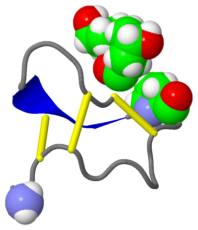
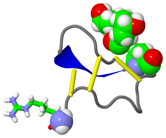
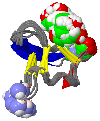
NMR StructureChain A from PDB Type:PROTEIN Length:25 aligned with CM3E_CONPU | P56529 from UniProtKB/Swiss-Prot Length:75 Alignment length:25 60 70 CM3E_CONPU 51 HPPCCMYGRCRRYPGCSSASCCQGG 75 SCOP domains d1as5a_ A: SCOP domains CATH domains ------------------------- CATH domains Pfam domains ------------------------- Pfam domains SAPs(SNPs) -----L--K--------------R- SAPs(SNPs) PROSITE ------------------------- PROSITE Transcript ------------------------- Transcript 1as5 A 1 HppCCLYGKCRRYpGCSSASCCQRx 25 || 10 | 20 | || 14-HYP 25-NH2 2-HYP 3-HYP
|
||||||||||||||||||||
SCOP Domains (1, 1)
NMR Structure
 |
CATH Domains (0, 0)| (no "CATH Domain" information available for 1AS5) |
Pfam Domains (0, 0)| (no "Pfam Domain" information available for 1AS5) |
Gene Ontology (4, 4)|
NMR Structure(hide GO term definitions) Chain A (CM3E_CONPU | P56529)
|
||||||||||||||||||||||||||||||||||||||||||
Interactive Views
|
|||||||||||||||||||||||||||||||||||||||||||||||||||||||||||||||||||||||||||||||||||||||||||||||||||||||||||||||||||||||||||||
Still Images
|
||||||||||||||||
Databases
|
||||||||||||||||||||||||||||||||||||||||||||||||||||||||||||||||||||||||||||||||||||||||||||||||||||||||||||||||||||||||||||||||||||||||||||||||||||||||||||||||
Analysis Tools
|
|||||||||||||||||||||||||||||||||||||||||||||||||||||||||||||
Entries Sharing at Least One Protein Chain (UniProt ID)
Related Entries Specified in the PDB File
|
|