| molecular function |
|---|
| | GO:0008017 | | microtubule binding | | Interacting selectively and non-covalently with microtubules, filaments composed of tubulin monomers. |
| | GO:0051010 | | microtubule plus-end binding | | Interacting selectively and non-covalently with the plus end of a microtubule. |
| | GO:0008022 | | protein C-terminus binding | | Interacting selectively and non-covalently with a protein C-terminus, the end of any peptide chain at which the 1-carboxy function of a constituent amino acid is not attached in peptide linkage to another amino-acid residue. |
| | GO:0005515 | | protein binding | | Interacting selectively and non-covalently with any protein or protein complex (a complex of two or more proteins that may include other nonprotein molecules). |
| biological process |
|---|
| | GO:0000086 | | G2/M transition of mitotic cell cycle | | The mitotic cell cycle transition by which a cell in G2 commits to M phase. The process begins when the kinase activity of M cyclin/CDK complex reaches a threshold high enough for the cell cycle to proceed. This is accomplished by activating a positive feedback loop that results in the accumulation of unphosphorylated and active M cyclin/CDK complex. |
| | GO:0007049 | | cell cycle | | The progression of biochemical and morphological phases and events that occur in a cell during successive cell replication or nuclear replication events. Canonically, the cell cycle comprises the replication and segregation of genetic material followed by the division of the cell, but in endocycles or syncytial cells nuclear replication or nuclear division may not be followed by cell division. |
| | GO:0051301 | | cell division | | The process resulting in division and partitioning of components of a cell to form more cells; may or may not be accompanied by the physical separation of a cell into distinct, individually membrane-bounded daughter cells. |
| | GO:0008283 | | cell proliferation | | The multiplication or reproduction of cells, resulting in the expansion of a cell population. |
| | GO:1904527 | | negative regulation of microtubule binding | | Any process that stops, prevents or reduces the frequency, rate or extent of microtubule binding. |
| | GO:0031115 | | negative regulation of microtubule polymerization | | Any process that stops, prevents, or reduces the frequency, rate or extent of microtubule polymerization. |
| | GO:0030335 | | positive regulation of cell migration | | Any process that activates or increases the frequency, rate or extent of cell migration. |
| | GO:1903033 | | positive regulation of microtubule plus-end binding | | Any process that activates or increases the frequency, rate or extent of microtubule plus-end binding. |
| | GO:0035372 | | protein localization to microtubule | | A process in which a protein is transported to, or maintained at, a microtubule. |
| | GO:0007062 | | sister chromatid cohesion | | The cell cycle process in which the sister chromatids of a replicated chromosome become tethered to each other. |
| cellular component |
|---|
| | GO:0005794 | | Golgi apparatus | | A compound membranous cytoplasmic organelle of eukaryotic cells, consisting of flattened, ribosome-free vesicles arranged in a more or less regular stack. The Golgi apparatus differs from the endoplasmic reticulum in often having slightly thicker membranes, appearing in sections as a characteristic shallow semicircle so that the convex side (cis or entry face) abuts the endoplasmic reticulum, secretory vesicles emerging from the concave side (trans or exit face). In vertebrate cells there is usually one such organelle, while in invertebrates and plants, where they are known usually as dictyosomes, there may be several scattered in the cytoplasm. The Golgi apparatus processes proteins produced on the ribosomes of the rough endoplasmic reticulum; such processing includes modification of the core oligosaccharides of glycoproteins, and the sorting and packaging of proteins for transport to a variety of cellular locations. Three different regions of the Golgi are now recognized both in terms of structure and function: cis, in the vicinity of the cis face, trans, in the vicinity of the trans face, and medial, lying between the cis and trans regions. |
| | GO:0042995 | | cell projection | | A prolongation or process extending from a cell, e.g. a flagellum or axon. |
| | GO:0031253 | | cell projection membrane | | The portion of the plasma membrane surrounding a plasma membrane bounded cell surface projection. |
| | GO:0005813 | | centrosome | | A structure comprised of a core structure (in most organisms, a pair of centrioles) and peripheral material from which a microtubule-based structure, such as a spindle apparatus, is organized. Centrosomes occur close to the nucleus during interphase in many eukaryotic cells, though in animal cells it changes continually during the cell-division cycle. |
| | GO:0030981 | | cortical microtubule cytoskeleton | | The portion of the microtubule cytoskeleton that lies just beneath the plasma membrane. |
| | GO:0005737 | | cytoplasm | | All of the contents of a cell excluding the plasma membrane and nucleus, but including other subcellular structures. |
| | GO:0005881 | | cytoplasmic microtubule | | Any microtubule in the cytoplasm of a cell. |
| | GO:0005856 | | cytoskeleton | | Any of the various filamentous elements that form the internal framework of cells, and typically remain after treatment of the cells with mild detergent to remove membrane constituents and soluble components of the cytoplasm. The term embraces intermediate filaments, microfilaments, microtubules, the microtrabecular lattice, and other structures characterized by a polymeric filamentous nature and long-range order within the cell. The various elements of the cytoskeleton not only serve in the maintenance of cellular shape but also have roles in other cellular functions, including cellular movement, cell division, endocytosis, and movement of organelles. |
| | GO:0005829 | | cytosol | | The part of the cytoplasm that does not contain organelles but which does contain other particulate matter, such as protein complexes. |
| | GO:0005874 | | microtubule | | Any of the long, generally straight, hollow tubes of internal diameter 12-15 nm and external diameter 24 nm found in a wide variety of eukaryotic cells; each consists (usually) of 13 protofilaments of polymeric tubulin, staggered in such a manner that the tubulin monomers are arranged in a helical pattern on the microtubular surface, and with the alpha/beta axes of the tubulin subunits parallel to the long axis of the tubule; exist in equilibrium with pool of tubulin monomers and can be rapidly assembled or disassembled in response to physiological stimuli; concerned with force generation, e.g. in the spindle. |
| | GO:0015630 | | microtubule cytoskeleton | | The part of the cytoskeleton (the internal framework of a cell) composed of microtubules and associated proteins. |
| | GO:0005815 | | microtubule organizing center | | An intracellular structure that can catalyze gamma-tubulin-dependent microtubule nucleation and that can anchor microtubules by interacting with their minus ends, plus ends or sides. |
| | GO:0035371 | | microtubule plus-end | | The growing (plus) end of a microtubule. In vitro, microtubules polymerize more quickly at the plus end than at the minus end. In vivo, microtubule growth occurs only at the plus end, and the plus end switches between periods of growth and shortening, a behavior known as dynamic instability. |
| | GO:0005819 | | spindle | | The array of microtubules and associated molecules that forms between opposite poles of a eukaryotic cell during mitosis or meiosis and serves to move the duplicated chromosomes apart. |
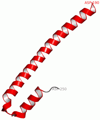
 Description
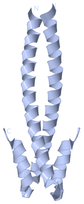