Show PDB file:   
         Plain Text   HTML   (compressed file size)
QuickSearch:   
by PDB,NDB,UniProt,PROSITE Code or Search Term(s)  
(-)NMR Structure - manually
(-)NMR Structure - model 1
(-)NMR Structure - model 1, sites
(-)NMR Structure - all models
collapse expand < >
Image NMR Structure - manually
NMR Structure - manually  (Jmol Viewer)
Image NMR Structure - model 1
NMR Structure - model 1  (Jmol Viewer)
Image NMR Structure - model 1, sites
NMR Structure - model 1, sites  (Jmol Viewer)
Image NMR Structure - all models
NMR Structure - all models  (Jmol Viewer)

(-) Description

Title :  THE SOLUTION STRUCTURE OF THE NISIN-LIPID II COMPLEX
 
Authors :  S. -T. D. Hsu, E. Breukink, E. Tischenko, M. A. G. Lutters, B. De Kruijff R. Kaptein, A. M. J. J. Bonvin, N. A. J. Van Nuland
Date :  19 Nov 04  (Deposition) - 07 Mar 05  (Release) - 13 Jul 11  (Revision)
Method :  SOLUTION NMR
Resolution :  NOT APPLICABLE
Chains :  NMR Structure  :  L,N  (20x)
NMR Structure *:  L,N  (1x)
Keywords :  Peptide-Antibiotic Complex, Lantibiotic, Antimicrobial, Bacteriocin, Thioester, Pore Formation, Pyrophosphate Cage, Food Preservative (Keyword Search: [Gene Ontology, PubMed, Web (Google))
 
Reference :  S. -T. D. Hsu, E. Breukink, E. Tischenko, M. A. G. Lutters, B. De Kruijff, R. Kaptein, A. M. J. J. Bonvin, N. A. J. Van Nuland
The Nisin-Lipid Ii Complex Reveals A Pyrophosphate Cage That Provides A Blueprint For Novel Antibiotics
Nat. Struct. Mol. Biol. V. 11 963 2004
PubMed-ID: 15361862  |  Reference-DOI: 10.1038/NSMB830
(for further references see the PDB file header)

(-) Compounds

Molecule 1 - NISIN Z
    ChainsN
    Organism ScientificLACTOCOCCUS LACTIS
    Organism Taxid1358
    StrainNIZO 22186
 
Molecule 2 - NAG-MUB-ALA-FGA-LYS-DAL-DAL PEPTIDE
    ChainsL
    Organism ScientificMONARTHROPALPUS FLAVUS
    Organism Taxid71816

 Structural Features

(-) Chains, Units

  12
NMR Structure (20x)LN
NMR Structure * (1x)LN

Summary Information (see also Sequences/Alignments below)

(-) Ligands, Modified Residues, Ions  (8, 14)

NMR Structure (8, 14)
No.NameCountTypeFull Name
1DAL3Mod. Amino AcidD-ALANINE
2DBB4Mod. Amino AcidD-ALPHA-AMINOBUTYRIC ACID
3DBU1Mod. Amino Acid(2E)-2-AMINOBUT-2-ENOIC ACID
4DHA2Mod. Amino Acid2-AMINO-ACRYLIC ACID
5FGA1Mod. Amino AcidD-GAMMA GLUTAMIC ACID
6FPP1Ligand/IonFARNESYL DIPHOSPHATE
7MUB1Mod. Amino AcidN-ACETYLMURAMIC ACID
8NAG1Mod. Amino AcidN-ACETYL-D-GLUCOSAMINE
NMR Structure * (8, 14)
No.NameCountTypeFull Name
1DAL3Mod. Amino AcidD-ALANINE
2DBB4Mod. Amino AcidD-ALPHA-AMINOBUTYRIC ACID
3DBU1Mod. Amino Acid(2E)-2-AMINOBUT-2-ENOIC ACID
4DHA2Mod. Amino Acid2-AMINO-ACRYLIC ACID
5FGA1Mod. Amino AcidD-GAMMA GLUTAMIC ACID
6FPP1Ligand/IonFARNESYL DIPHOSPHATE
7MUB1Mod. Amino AcidN-ACETYLMURAMIC ACID
8NAG1Mod. Amino AcidN-ACETYL-D-GLUCOSAMINE

(-) Sites  (3, 3)

NMR Structure (3, 3)
No.NameEvidenceResiduesDescription
1AC1SOFTWAREMUB L:2 , ILE N:1 , DBU N:2 , DAL N:3 , ILE N:4 , DHA N:5 , LEU N:6 , CYS N:7 , DBB N:8 , PRO N:9 , GLY N:10BINDING SITE FOR RESIDUE FPP N 35
2AC2SOFTWAREMUB L:2 , ALA L:3 , FGA L:4 , DAL L:6 , DAL L:7 , FPP N:35BINDING SITE FOR CHAIN N OF NISIN Z
3AC3SOFTWAREILE N:1 , DBU N:2 , ILE N:4 , GLY N:10 , DBB N:13 , GLY N:14 , FPP N:35BINDING SITE FOR CHAIN L OF NAG-MUB-ALA-FGA-LYS-DAL-DAL PEPTIDE

(-) SS Bonds  (0, 0)

(no "SS Bond" information available for 1WCO)

(-) Cis Peptide Bonds  (0, 0)

(no "Cis Peptide Bond" information available for 1WCO)

 Sequence-Structure Mapping

(-) SAPs(SNPs)/Variants  (1, 1)

NMR Structure (1, 1)
  dbSNPPDB
No.SourceVariant IDVariantUniProt IDStatusIDChainVariant
1UniProtVAR_LANZ_LACLL_001 *N50HLANZ_LACLL  ---  ---NN27H
   * ID not provided by source

  SNP/SAP Summary Statistics (UniProtKB/Swiss-Prot)
NMR Structure * (1, 1)
  dbSNPPDB
No.SourceVariant IDVariantUniProt IDStatusIDChainVariant
1UniProtVAR_LANZ_LACLL_001 *N50HLANZ_LACLL  ---  ---NN27H
   * ID not provided by source

  SNP/SAP Summary Statistics (UniProtKB/Swiss-Prot)

(-) PROSITE Motifs  (0, 0)

(no "PROSITE Motif" information available for 1WCO)

(-) Exons   (0, 0)

(no "Exon" information available for 1WCO)

(-) Sequences/Alignments

NMR Structure
   Reformat: Number of residues per line =  ('0' or empty: single-line sequence representation)
  Number of residues per labelling interval =   
  UniProt sequence: complete  aligned part    
   Show mapping: SCOP domains CATH domains Pfam domains Secondary structure (by author)
SAPs(SNPs) PROSITE motifs Exons
(details for a mapped element are shown in a popup box when the mouse pointer rests over it)
Chain L from PDB  Type:PROTEIN  Length:7
                                      
               SCOP domains ------- SCOP domains
               CATH domains ------- CATH domains
               Pfam domains ------- Pfam domains
         Sec.struct. author ....... Sec.struct. author
                 SAPs(SNPs) ------- SAPs(SNPs)
                    PROSITE ------- PROSITE
                 Transcript ------- Transcript
                  1wco L  1 xxAxKss  7
                            || | ||
                            || | ||
                            1-NAG||
                             2-MUB|
                               4-FGA
                                 6-DAL
                                  7-DAL

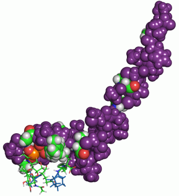
Chain N from PDB  Type:PROTEIN  Length:34
 aligned with LANZ_LACLL | P29559 from UniProtKB/Swiss-Prot  Length:57

    Alignment length:34
                                    33        43        53    
            LANZ_LACLL   24 ITSISLCTPGCKTGALMGCNMKTATCNCSIHVSK 57
               SCOP domains d1wcon1 N:1-34 Nisin               SCOP domains
               CATH domains ---------------------------------- CATH domains
               Pfam domains ---------------------------------- Pfam domains
         Sec.struct. author .......hhhhhh..................... Sec.struct. author
                 SAPs(SNPs) --------------------------H------- SAPs(SNPs)
                    PROSITE ---------------------------------- PROSITE
                 Transcript ---------------------------------- Transcript
                  1wco N  1 ItsIsLCtPGCKtGALMGCNMKtAtCNCSIHVsK 34
                             || |  |10  |     20  | |   30  | 
                             || |  |    |        23-DBB    33-DHA
                             2-DBU |    |          25-DBB     
                              3-DAL|    |                     
                                5-DHA   |                     
                                   8-DBB|                     
                                       13-DBB                 

   Legend:   → Mismatch (orange background)
  - → Gap (green background, '-', border residues have a numbering label)
    → Modified Residue (blue background, lower-case, 'x' indicates undefined single-letter code, labelled with number + name)
  x → Chemical Group (purple background, 'x', labelled with number + name, e.g. ACE or NH2)
  extra numbering lines below/above indicate numbering irregularities and modified residue names etc., number ends below/above '|'

 Classification and Annotation

(-) SCOP Domains  (1, 1)

NMR Structure
(-)
Class: Peptides (792)

(-) CATH Domains  (0, 0)

(no "CATH Domain" information available for 1WCO)

(-) Pfam Domains  (0, 0)

(no "Pfam Domain" information available for 1WCO)

(-) Gene Ontology  (4, 4)

NMR Structure(hide GO term definitions)
Chain N   (LANZ_LACLL | P29559)
molecular function
    GO:0005102    receptor binding    Interacting selectively and non-covalently with one or more specific sites on a receptor molecule, a macromolecule that undergoes combination with a hormone, neurotransmitter, drug or intracellular messenger to initiate a change in cell function.
biological process
    GO:0019835    cytolysis    The rupture of cell membranes and the loss of cytoplasm.
    GO:0042742    defense response to bacterium    Reactions triggered in response to the presence of a bacterium that act to protect the cell or organism.
cellular component
    GO:0005576    extracellular region    The space external to the outermost structure of a cell. For cells without external protective or external encapsulating structures this refers to space outside of the plasma membrane. This term covers the host cell environment outside an intracellular parasite.

 Visualization

(-) Interactive Views

NMR Structure
  Complete Structure
    Jena3D(integrated viewing of ligand, site, SAP, PROSITE, SCOP information)
    WebMol | AstexViewer[tm]@PDBe
(Java Applets, require no local installation except for Java; loading may be slow)
    STRAP
(Java WebStart application, automatic local installation, requires Java; full application with system access!)
    RasMol
(require local installation)
    Molscript (VRML)
(requires installation of a VRML viewer; select preferred view via VRML and generate a mono or stereo PDF format file)
 
  Ligands, Modified Residues, Ions
    DAL  [ RasMol | Jena3D ]  +environment [ RasMol | Jena3D ]
    DBB  [ RasMol | Jena3D ]  +environment [ RasMol | Jena3D ]
    DBU  [ RasMol | Jena3D ]  +environment [ RasMol | Jena3D ]
    DHA  [ RasMol | Jena3D ]  +environment [ RasMol | Jena3D ]
    FGA  [ RasMol | Jena3D ]  +environment [ RasMol | Jena3D ]
    FPP  [ RasMol | Jena3D ]  +environment [ RasMol | Jena3D ]
    MUB  [ RasMol | Jena3D ]  +environment [ RasMol | Jena3D ]
    NAG  [ RasMol | Jena3D ]  +environment [ RasMol | Jena3D ]
 
  Sites
    AC1  [ RasMol ]  +environment [ RasMol ]
    AC2  [ RasMol ]  +environment [ RasMol ]
    AC3  [ RasMol ]  +environment [ RasMol ]
 
  Cis Peptide Bonds
(no "Cis Peptide Bonds" information available for 1wco)
 

(-) Still Images

Jmol
  protein: cartoon or spacefill or dots and stick; nucleic acid: cartoon and stick; ligands: spacefill; active site: stick
Molscript
  protein, nucleic acid: cartoon; ligands: spacefill; active site: ball and stick

 Databases and Analysis Tools

(-) Databases

Access by PDB/NDB ID
  1wco
    Family and Domain InformationProDom | SYSTERS
    General Structural InformationGlycoscienceDB | MMDB | NDB | OCA | PDB | PDBe | PDBj | PDBsum | PDBWiki | PQS | PROTEOPEDIA
    Orientation in MembranesOPM
    Protein SurfaceSURFACE
    Secondary StructureDSSP (structure derived) | HSSP (homology derived)
    Structural GenomicsGeneCensus
    Structural NeighboursCE | VAST
    Structure ClassificationCATH | Dali | SCOP
    Validation and Original DataBMRB Data View | BMRB Restraints Grid | EDS | PROCHECK | RECOORD | WHAT_CHECK
 
Access by UniProt ID/Accession number
  LANZ_LACLL | P29559
    Comparative Protein Structure ModelsModBase
    Genomic InformationEnsembl
    Protein-protein InteractionDIP
    Sequence, Family and Domain InformationInterPro | Pfam | SMART | UniProtKB/SwissProt
 
Access by Enzyme Classificator   (EC Number)
  (no 'Enzyme Classificator' available)
    General Enzyme InformationBRENDA | EC-PDB | Enzyme | IntEnz
    PathwayKEGG | MetaCyc
 
Access by Disease Identifier   (MIM ID)
  (no 'MIM ID' available)
    Disease InformationOMIM
 
Access by GenAge ID
  (no 'GenAge ID' available)
    Age Related InformationGenAge

(-) Analysis Tools

Access by PDB/NDB ID
    Domain InformationXDom
    Interatomic Contacts of Structural UnitsCSU
    Ligand-protein ContactsLPC
    Protein CavitiescastP
    Sequence and Secondary StructurePDBCartoon
    Structure AlignmentSTRAP(Java WebStart application, automatic local installation, requires Java; full application with system access!)
    Structure and Sequence BrowserSTING
 
Access by UniProt ID/Accession number
  LANZ_LACLL | P29559
    Protein Disorder PredictionDisEMBL | FoldIndex | GLOBPLOT (for more information see DisProt)

 Related Entries

(-) Entries Sharing at Least One Protein Chain (UniProt ID)

(no "Entries Sharing at Least One Protein Chain" available for 1WCO)

(-) Related Entries Specified in the PDB File

1aj1 SOLUTION STRUCTURE OF THE LANTIBIOTIC ACTAGARDINE
1mqx SOLUTION STRUCTURE OF TYPE B LANTIBIOTICS MERSACIDIN IN MEOH/H2O MIXTURE
1mqy SOLUTION STRUCTURE OF TYPE B LANTIBIOTICS MERSACIDIN IN DPC MICELLES
1mqz SOLUTION STRUCTURE OF TYPE B LANTIBIOTICS MERSACIDIN BOUND TO LIPID II IN DPC MICELLES
1qow CRYSTAL STRUCTURE OF THE TUPE B LANTIBIOTIC MERSACIDIN
1w9n SOLUTION STRUCTURE OF THE LANTIBIOTIC EPILANCIN 15X
2dde SOLUTION STRUCTURE OF THE LANTIBIOTIC CINNAMYCIN COMPLEXED WITH LYSOPHOSPHATIDYLETHANOLAMINE
2ktn SOLUTION STRUCTURE OF LCH-ALPHA PEPTIDE FROM TWO-COMPONENT LANTIBIOTIC SYSTEM LICHENICIDIN VK21 A1
2kto SOLUTION STRUCTURE OF LCH-BETA PEPTIDE FROM TWO-COMPONENT LANTIBIOTIC LICHENICIDIN VK21 A2