| molecular function |
|---|
| | GO:0019838 | | growth factor binding | | Interacting selectively and non-covalently with any growth factor, proteins or polypeptides that stimulate a cell or organism to grow or proliferate. |
| | GO:0031994 | | insulin-like growth factor I binding | | Interacting selectively and non-covalently with insulin-like growth factor I. |
| | GO:0031995 | | insulin-like growth factor II binding | | Interacting selectively and non-covalently with insulin-like growth factor II. |
| | GO:0005520 | | insulin-like growth factor binding | | Interacting selectively and non-covalently with an insulin-like growth factor, any member of a group of polypeptides that are structurally homologous to insulin and share many of its biological activities, but are immunologically distinct from it. |
| | GO:0005102 | | receptor binding | | Interacting selectively and non-covalently with one or more specific sites on a receptor molecule, a macromolecule that undergoes combination with a hormone, neurotransmitter, drug or intracellular messenger to initiate a change in cell function. |
| biological process |
|---|
| | GO:0044267 | | cellular protein metabolic process | | The chemical reactions and pathways involving a specific protein, rather than of proteins in general, occurring at the level of an individual cell. Includes cellular protein modification. |
| | GO:0090090 | | negative regulation of canonical Wnt signaling pathway | | Any process that decreases the rate, frequency, or extent of the Wnt signaling pathway through beta-catenin, the series of molecular signals initiated by binding of a Wnt protein to a frizzled family receptor on the surface of the target cell, followed by propagation of the signal via beta-catenin, and ending with a change in transcription of target genes. |
| | GO:0008285 | | negative regulation of cell proliferation | | Any process that stops, prevents or reduces the rate or extent of cell proliferation. |
| | GO:0001558 | | regulation of cell growth | | Any process that modulates the frequency, rate, extent or direction of cell growth. |
| | GO:0043567 | | regulation of insulin-like growth factor receptor signaling pathway | | Any process that modulates the frequency, rate or extent of insulin-like growth factor receptor signaling. |
| | GO:0007165 | | signal transduction | | The cellular process in which a signal is conveyed to trigger a change in the activity or state of a cell. Signal transduction begins with reception of a signal (e.g. a ligand binding to a receptor or receptor activation by a stimulus such as light), or for signal transduction in the absence of ligand, signal-withdrawal or the activity of a constitutively active receptor. Signal transduction ends with regulation of a downstream cellular process, e.g. regulation of transcription or regulation of a metabolic process. Signal transduction covers signaling from receptors located on the surface of the cell and signaling via molecules located within the cell. For signaling between cells, signal transduction is restricted to events at and within the receiving cell. |
| cellular component |
|---|
| | GO:0005794 | | Golgi apparatus | | A compound membranous cytoplasmic organelle of eukaryotic cells, consisting of flattened, ribosome-free vesicles arranged in a more or less regular stack. The Golgi apparatus differs from the endoplasmic reticulum in often having slightly thicker membranes, appearing in sections as a characteristic shallow semicircle so that the convex side (cis or entry face) abuts the endoplasmic reticulum, secretory vesicles emerging from the concave side (trans or exit face). In vertebrate cells there is usually one such organelle, while in invertebrates and plants, where they are known usually as dictyosomes, there may be several scattered in the cytoplasm. The Golgi apparatus processes proteins produced on the ribosomes of the rough endoplasmic reticulum; such processing includes modification of the core oligosaccharides of glycoproteins, and the sorting and packaging of proteins for transport to a variety of cellular locations. Three different regions of the Golgi are now recognized both in terms of structure and function: cis, in the vicinity of the cis face, trans, in the vicinity of the trans face, and medial, lying between the cis and trans regions. |
| | GO:0005737 | | cytoplasm | | All of the contents of a cell excluding the plasma membrane and nucleus, but including other subcellular structures. |
| | GO:0070062 | | extracellular exosome | | A vesicle that is released into the extracellular region by fusion of the limiting endosomal membrane of a multivesicular body with the plasma membrane. Extracellular exosomes, also simply called exosomes, have a diameter of about 40-100 nm. |
| | GO:0005576 | | extracellular region | | The space external to the outermost structure of a cell. For cells without external protective or external encapsulating structures this refers to space outside of the plasma membrane. This term covers the host cell environment outside an intracellular parasite. |
| | GO:0005615 | | extracellular space | | That part of a multicellular organism outside the cells proper, usually taken to be outside the plasma membranes, and occupied by fluid. |
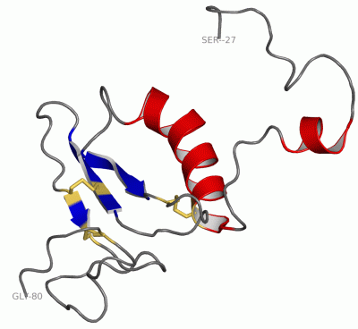
 Description
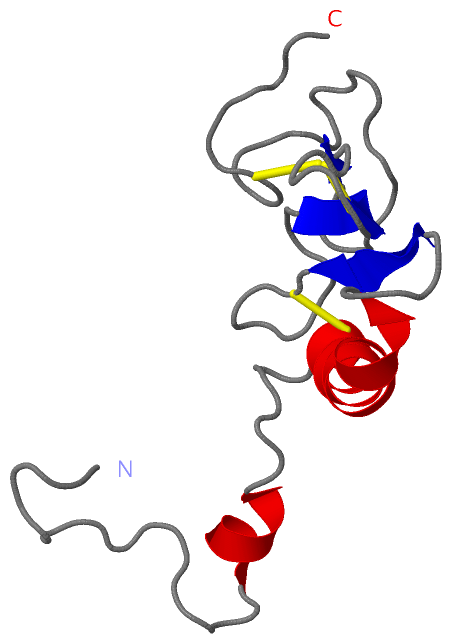