|
|
|
|
 Description
|
|
Compounds
|
||||||||||||||||||||||||||||||||||||||||||||||||||||
Chains, Units
Summary Information (see also Sequences/Alignments below) |
Ligands, Modified Residues, Ions (3, 6)
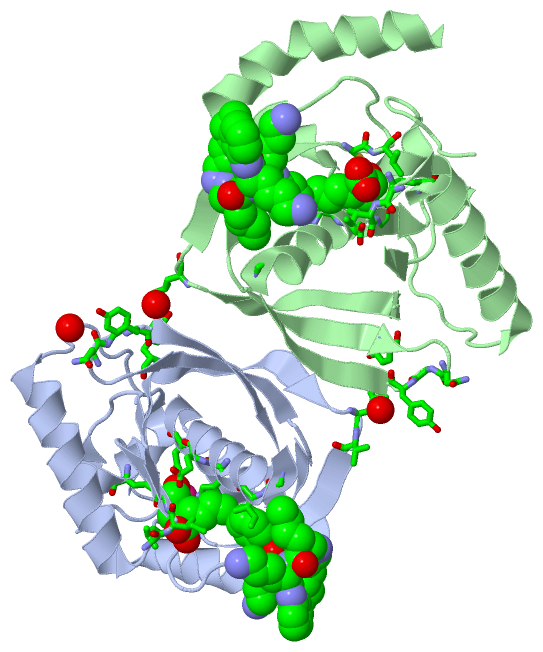
Asymmetric Unit (3, 6)
|
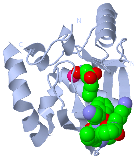
Sites (6, 6)
Asymmetric Unit (6, 6)
|
SS Bonds (0, 0)| (no "SS Bond" information available for 1RL4) |
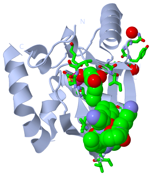
Cis Peptide Bonds (2, 2)
Asymmetric Unit
|
||||||||||||
SAPs(SNPs)/Variants (0, 0)| (no "SAP(SNP)/Variant" information available for 1RL4) |
PROSITE Motifs (0, 0)| (no "PROSITE Motif" information available for 1RL4) |
Exons (0, 0)| (no "Exon" information available for 1RL4) |
Sequences/Alignments
Asymmetric UnitChain A from PDB Type:PROTEIN Length:154 aligned with Q8I372_PLAF7 | Q8I372 from UniProtKB/TrEMBL Length:241 Alignment length:165 75 85 95 105 115 125 135 145 155 165 175 185 195 205 215 225 Q8I372_PLAF7 66 KIVKYPDPILRRRSEEVTNFDDNLKRVVRKMFDIMYESKGIGLSAPQVNISKRIIVWNALYEKRKEENERIFINPSIVEQSLVKLKLIEGCLSFPGIEGKVERPSIVSISYYDINGYKHLKILKGIHSRIFQHEFDHLNGTLFIDKMTQVDKKKVRPKLNELIRD 230 SCOP domains d1rl4a_ A: Peptide deformylase SCOP domains CATH domains 1rl4A00 A:66-230 Peptide Deformylase CATH domains Pfam domains --------------------------------------------------------------------------------------------------------------------------------------------------------------------- Pfam domains SAPs(SNPs) --------------------------------------------------------------------------------------------------------------------------------------------------------------------- SAPs(SNPs) PROSITE --------------------------------------------------------------------------------------------------------------------------------------------------------------------- PROSITE Transcript --------------------------------------------------------------------------------------------------------------------------------------------------------------------- Transcript 1rl4 A 66 KIVKYPDPILRRRSEEVTNFDDNLKRVVRKMFDIMYESKGIGLSAPQVNISKRIIVWN-----------RIFINPSIVEQSLVKLKLIEGCLSFPGIEGKVERPSIVSISYYDINGYKHLKILKGIHSRIFQHEFDHLNGTLFIDKMTQVDKKKVRPKLNELIRD 230 75 85 95 105 115 | - 135 145 155 165 175 185 195 205 215 225 123 135 Chain B from PDB Type:PROTEIN Length:156 aligned with Q8I372_PLAF7 | Q8I372 from UniProtKB/TrEMBL Length:241 Alignment length:167 75 85 95 105 115 125 135 145 155 165 175 185 195 205 215 225 Q8I372_PLAF7 66 KIVKYPDPILRRRSEEVTNFDDNLKRVVRKMFDIMYESKGIGLSAPQVNISKRIIVWNALYEKRKEENERIFINPSIVEQSLVKLKLIEGCLSFPGIEGKVERPSIVSISYYDINGYKHLKILKGIHSRIFQHEFDHLNGTLFIDKMTQVDKKKVRPKLNELIRDYK 232 SCOP domains d1rl4b_ B: Peptide deformylase SCOP domains CATH domains 1rl4B00 B:66-232 Peptide Deformylase CATH domains Pfam domains (1) Pep_deformylase-1rl4B01 B:66-218 -------------- Pfam domains (1) Pfam domains (2) Pep_deformylase-1rl4B02 B:66-218 -------------- Pfam domains (2) SAPs(SNPs) ----------------------------------------------------------------------------------------------------------------------------------------------------------------------- SAPs(SNPs) PROSITE ----------------------------------------------------------------------------------------------------------------------------------------------------------------------- PROSITE Transcript ----------------------------------------------------------------------------------------------------------------------------------------------------------------------- Transcript 1rl4 B 66 KIVKYPDPILRRRSEEVTNFDDNLKRVVRKMFDIMYESKGIGLSAPQVNISKRIIVWN-----------RIFINPSIVEQSLVKLKLIEGCLSFPGIEGKVERPSIVSISYYDINGYKHLKILKGIHSRIFQHEFDHLNGTLFIDKMTQVDKKKVRPKLNELIRDYK 232 75 85 95 105 115 | - 135 145 155 165 175 185 195 205 215 225 123 135
|
||||||||||||||||||||
SCOP Domains (1, 2)
Asymmetric Unit
 |
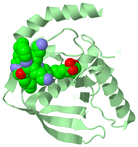
CATH Domains (1, 2)
Asymmetric Unit
|
Pfam Domains (1, 2)
Asymmetric Unit
|
Gene Ontology (11, 11)|
Asymmetric Unit(hide GO term definitions) Chain A,B (Q8I372_PLAF7 | Q8I372)
|
||||||||||||||||||||||||||||||||||||||||||||||||||||||||||||||||||||||||||||||||||||
Interactive Views
|
||||||||||||||||||||||||||||||||||||||||||||||||||||||||||||||||||||||||||||||||||||||||||||||||||||||||||||||||||||||||||||||||||||||||||||||||||||||||||||||||||||||||||||||||||||||||||||||||||||||
Still Images
|
||||||||||||||||
Databases
|
||||||||||||||||||||||||||||||||||||||||||||||||||||||||||||||||||||||||||||||||||||||||||||||||||||||||||||||||||||||||||||||||||||||||||||||||||||||||||||||||
Analysis Tools
|
|||||||||||||||||||||||||||||||||||||||||||||||||||||||||||||
Entries Sharing at Least One Protein Chain (UniProt ID)
Related Entries Specified in the PDB File
|
|