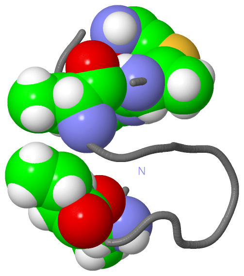
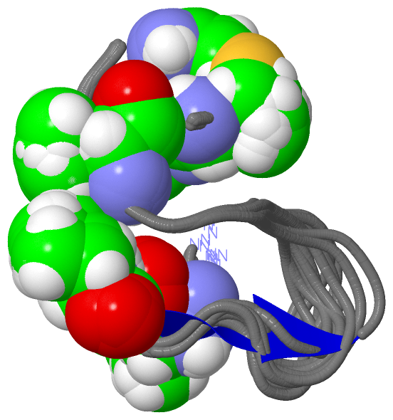
Show PDB file:   
         Plain Text   HTML   (compressed file size)
QuickSearch:   
by PDB,NDB,UniProt,PROSITE Code or Search Term(s)  
(-)NMR Structure - model 1
(-)NMR Structure - all models
collapse expand < >
Image NMR Structure - model 1
NMR Structure - model 1  (Jmol Viewer)
Image NMR Structure - all models
NMR Structure - all models  (Jmol Viewer)

(-) Description

Title :  NMR SOLUTION STRUCTURE OF TYPE-B LANTIBIOTICS MERSACIDIN IN DPC MICELLES
 
Authors :  S. -T. Hsu, E. Breukink, G. Bierbaum, H. -G. Sahl, B. De Kruijff, R. Kapt N. A. Van Nuland, A. M. Bonvin
Date :  17 Sep 02  (Deposition) - 11 Mar 03  (Release) - 27 Jul 11  (Revision)
Method :  SOLUTION NMR
Resolution :  NOT APPLICABLE
Chains :  NMR Structure  :  A  (13x)
NMR Structure *:  A  (1x)
Keywords :  Antibiotic, Antimicrobial, Lantibiotics, Bacteriocin, Peptidoglycan Methicillin Resistance, Thioester (Keyword Search: [Gene Ontology, PubMed, Web (Google))
 
Reference :  S. -T. Hsu, E. Breukink, G. Bierbaum, H. -G. Sahl, B. De Kruijff, R. Kaptein, N. A. Van Nuland, A. M. Bonvin
Nmr Study Of Mersacidin And Lipid Ii Interaction In Dodecylphosphocholine Micelles. Conformational Changes Are Key To Antimicrobial Activity
J. Biol. Chem. V. 278 13110 2003
PubMed-ID: 12562773  |  Reference-DOI: 10.1074/JBC.M211144200

(-) Compounds

Molecule 1 - LANTIBIOTIC MERSACIDIN
    ChainsA
    Organism ScientificBACILLUS SP. HIL-Y85/54728
    Organism Taxid69002
    StrainHIL Y-85, 54728
    SynonymTYPE-B LANTIBIOTICS MERSACIDIN

 Structural Features

(-) Chains, Units

  1
NMR Structure (13x)A
NMR Structure * (1x)A

Summary Information (see also Sequences/Alignments below)

(-) Ligands, Modified Residues, Ions  (3, 6)

NMR Structure (3, 6)
No.NameCountTypeFull Name
1DBB4Mod. Amino AcidD-ALPHA-AMINOBUTYRIC ACID
2DHA1Mod. Amino Acid2-AMINO-ACRYLIC ACID
3TEE1Mod. Amino Acid2-AMINO-ETHENETHIOL
NMR Structure * (3, 6)
No.NameCountTypeFull Name
1DBB4Mod. Amino AcidD-ALPHA-AMINOBUTYRIC ACID
2DHA1Mod. Amino Acid2-AMINO-ACRYLIC ACID
3TEE1Mod. Amino Acid2-AMINO-ETHENETHIOL

(-) Sites  (0, 0)

(no "Site" information available for 1MQY)

(-) SS Bonds  (0, 0)

(no "SS Bond" information available for 1MQY)

(-) Cis Peptide Bonds  (1, 13)

NMR Structure
No.ModelResidues
11, 2, 3, 4, 5, 6, 7, 8, 9, 10, 11, 12, 13Cys A:1 -Dbb A:2

 Sequence-Structure Mapping

(-) SAPs(SNPs)/Variants  (0, 0)

(no "SAP(SNP)/Variant" information available for 1MQY)

(-) PROSITE Motifs  (0, 0)

(no "PROSITE Motif" information available for 1MQY)

(-) Exons   (0, 0)

(no "Exon" information available for 1MQY)

(-) Sequences/Alignments

NMR Structure
   Reformat: Number of residues per line =  ('0' or empty: single-line sequence representation)
  Number of residues per labelling interval =   
  UniProt sequence: complete  aligned part    
   Show mapping: SCOP domains CATH domains Pfam domains Secondary structure (by author)
SAPs(SNPs) PROSITE motifs Exons
(details for a mapped element are shown in a popup box when the mouse pointer rests over it)
Chain A from PDB  Type:PROTEIN  Length:20
 aligned with MRSA_BACSY | P43683 from UniProtKB/Swiss-Prot  Length:68

    Alignment length:20
                                    58        68
            MRSA_BACSY   49 CTFTLPGGGGVCTLTSECIC 68
               SCOP domains d1mqya_ A:           SCOP domains
               CATH domains -------------------- CATH domains
               Pfam domains -------------------- Pfam domains
         Sec.struct. author .................... Sec.struct. author
                 SAPs(SNPs) -------------------- SAPs(SNPs)
                    PROSITE -------------------- PROSITE
                 Transcript -------------------- Transcript
                  1mqy A  1 CtFtLPGGGGVCtLtsECIc 20
                             | |    10  | ||  20
                             | |       13-DBB  |
                             2-DBB       15-DBB|
                               4-DBB      16-DHA
                                              20-TEE

   Legend:   → Mismatch (orange background)
  - → Gap (green background, '-', border residues have a numbering label)
    → Modified Residue (blue background, lower-case, 'x' indicates undefined single-letter code, labelled with number + name)
  x → Chemical Group (purple background, 'x', labelled with number + name, e.g. ACE or NH2)
  extra numbering lines below/above indicate numbering irregularities and modified residue names etc., number ends below/above '|'

 Classification and Annotation

(-) SCOP Domains  (1, 1)

NMR Structure
(-)
Class: Peptides (792)

(-) CATH Domains  (0, 0)

(no "CATH Domain" information available for 1MQY)

(-) Pfam Domains  (0, 0)

(no "Pfam Domain" information available for 1MQY)

(-) Gene Ontology  (3, 3)

NMR Structure(hide GO term definitions)
Chain A   (MRSA_BACSY | P43683)
molecular function
    GO:0005102    receptor binding    Interacting selectively and non-covalently with one or more specific sites on a receptor molecule, a macromolecule that undergoes combination with a hormone, neurotransmitter, drug or intracellular messenger to initiate a change in cell function.
biological process
    GO:0019835    cytolysis    The rupture of cell membranes and the loss of cytoplasm.
    GO:0042742    defense response to bacterium    Reactions triggered in response to the presence of a bacterium that act to protect the cell or organism.

 Visualization

(-) Interactive Views

NMR Structure
  Complete Structure
    Jena3D(integrated viewing of ligand, site, SAP, PROSITE, SCOP information)
    WebMol | AstexViewer[tm]@PDBe
(Java Applets, require no local installation except for Java; loading may be slow)
    STRAP
(Java WebStart application, automatic local installation, requires Java; full application with system access!)
    RasMol
(require local installation)
    Molscript (VRML)
(requires installation of a VRML viewer; select preferred view via VRML and generate a mono or stereo PDF format file)
 
  Ligands, Modified Residues, Ions
    DBB  [ RasMol | Jena3D ]  +environment [ RasMol | Jena3D ]
    DHA  [ RasMol | Jena3D ]  +environment [ RasMol | Jena3D ]
    TEE  [ RasMol | Jena3D ]  +environment [ RasMol | Jena3D ]
 
  Sites
(no "Sites" information available for 1mqy)
 
  Cis Peptide Bonds
    Cys A:1 - Dbb A:2   [ RasMol ]  
 

(-) Still Images

Jmol
  protein: cartoon or spacefill or dots and stick; nucleic acid: cartoon and stick; ligands: spacefill; active site: stick
Molscript
  protein, nucleic acid: cartoon; ligands: spacefill; active site: ball and stick

 Databases and Analysis Tools

(-) Databases

Access by PDB/NDB ID
  1mqy
    Family and Domain InformationProDom | SYSTERS
    General Structural InformationGlycoscienceDB | MMDB | NDB | OCA | PDB | PDBe | PDBj | PDBsum | PDBWiki | PQS | PROTEOPEDIA
    Orientation in MembranesOPM
    Protein SurfaceSURFACE
    Secondary StructureDSSP (structure derived) | HSSP (homology derived)
    Structural GenomicsGeneCensus
    Structural NeighboursCE | VAST
    Structure ClassificationCATH | Dali | SCOP
    Validation and Original DataBMRB Data View | BMRB Restraints Grid | EDS | PROCHECK | RECOORD | WHAT_CHECK
 
Access by UniProt ID/Accession number
  MRSA_BACSY | P43683
    Comparative Protein Structure ModelsModBase
    Genomic InformationEnsembl
    Protein-protein InteractionDIP
    Sequence, Family and Domain InformationInterPro | Pfam | SMART | UniProtKB/SwissProt
 
Access by Enzyme Classificator   (EC Number)
  (no 'Enzyme Classificator' available)
    General Enzyme InformationBRENDA | EC-PDB | Enzyme | IntEnz
    PathwayKEGG | MetaCyc
 
Access by Disease Identifier   (MIM ID)
  (no 'MIM ID' available)
    Disease InformationOMIM
 
Access by GenAge ID
  (no 'GenAge ID' available)
    Age Related InformationGenAge

(-) Analysis Tools

Access by PDB/NDB ID
    Domain InformationXDom
    Interatomic Contacts of Structural UnitsCSU
    Ligand-protein ContactsLPC
    Protein CavitiescastP
    Sequence and Secondary StructurePDBCartoon
    Structure AlignmentSTRAP(Java WebStart application, automatic local installation, requires Java; full application with system access!)
    Structure and Sequence BrowserSTING
 
Access by UniProt ID/Accession number
  MRSA_BACSY | P43683
    Protein Disorder PredictionDisEMBL | FoldIndex | GLOBPLOT (for more information see DisProt)

 Related Entries

(-) Entries Sharing at Least One Protein Chain (UniProt ID)

UniProtKB/Swiss-Prot
        MRSA_BACSY | P436831mqx 1mqz 1qow

(-) Related Entries Specified in the PDB File

1aj1 SOLUTION STRUCTURE OF THE LANTIBIOTIC ACTAGARDINE
1mqx SOLUTION STRUCTURE OF TYPE B LANTIBIOTICS MERSACIDIN IN MEOH/H2O MIXTURE
1mqz SOLUTION STRUCTURE OF TYPE B LANTIBIOTICS MERSACIDIN BOUND TO LIPID II IN DPC MICELLES
1qow CRYSTAL STRUCTURE OF THE TUPE B LANTIBIOTIC MERSACIDIN
1w9n SOLUTION STRUCTURE OF THE LANTIBIOTIC EPILANCIN 15X
1wco SOLUTION STRUCTURE OF NISIN AND LIPID II COMPLEX
2dde SOLUTION STRUCTURE OF THE LANTIBIOTIC CINNAMYCIN COMPLEXED WITH LYSOPHOSPHATIDYLETHANOLAMINE
2ktn SOLUTION STRUCTURE OF LCH-ALPHA PEPTIDE FROM TWO-COMPONENT LANTIBIOTIC SYSTEM LICHENICIDIN VK21 A1
2kto SOLUTION STRUCTURE OF LCH-BETA PEPTIDE FROM TWO-COMPONENT LANTIBIOTIC LICHENICIDIN VK21 A2