|
|
|
|
 Description
|
|
Compounds
|
||||||||||||||||||||||||||||||||
Chains, Units
Summary Information (see also Sequences/Alignments below) |
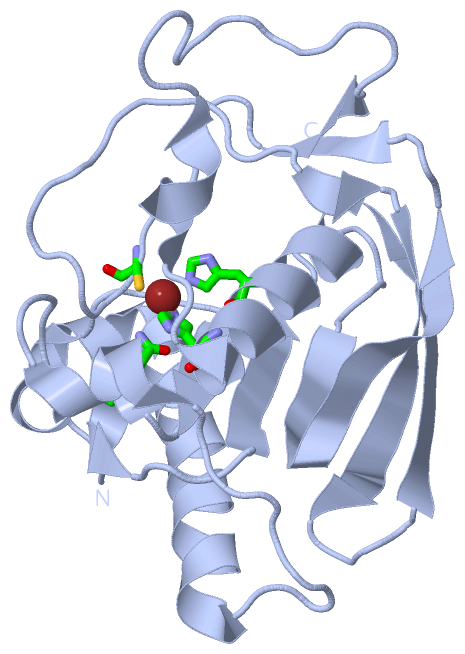
Ligands, Modified Residues, Ions (1, 2)
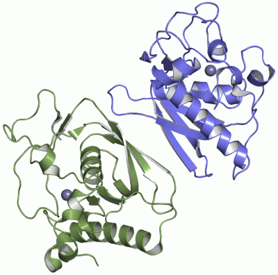
Asymmetric Unit (1, 2)
|
Sites (2, 2)
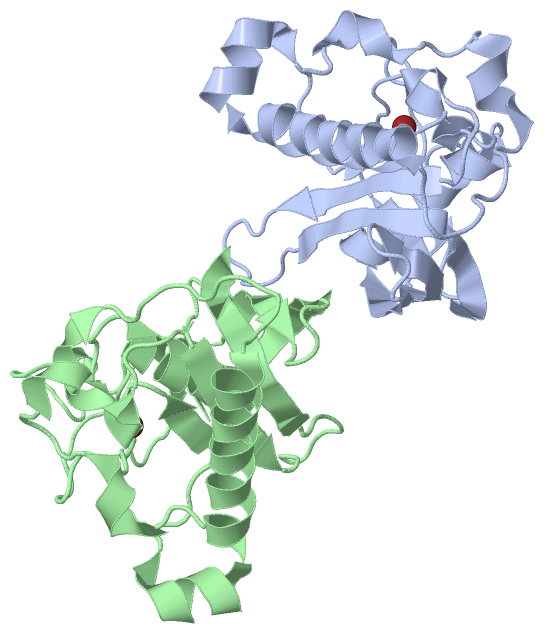
Asymmetric Unit (2, 2)
|
SS Bonds (0, 0)| (no "SS Bond" information available for 1LQW) |
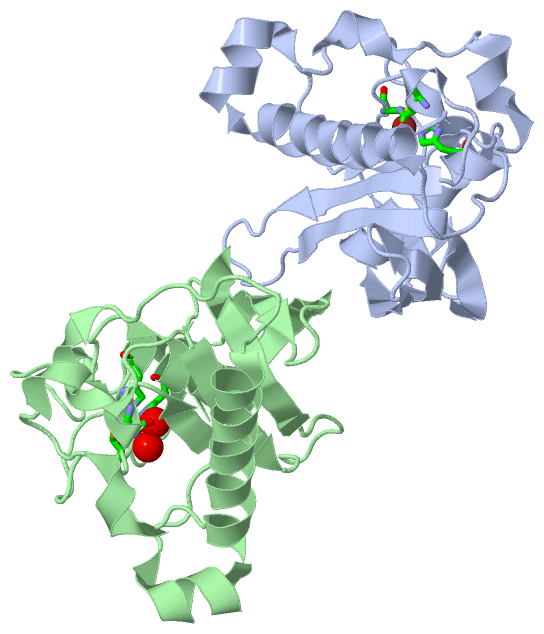
Cis Peptide Bonds (2, 2)
Asymmetric Unit
|
||||||||||||
SAPs(SNPs)/Variants (0, 0)| (no "SAP(SNP)/Variant" information available for 1LQW) |
PROSITE Motifs (0, 0)| (no "PROSITE Motif" information available for 1LQW) |
Exons (0, 0)| (no "Exon" information available for 1LQW) |
Sequences/Alignments
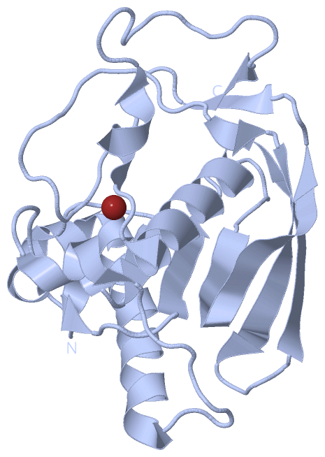
Asymmetric UnitChain A from PDB Type:PROTEIN Length:183 aligned with DEF_STAAU | P68826 from UniProtKB/Swiss-Prot Length:183 Alignment length:183 10 20 30 40 50 60 70 80 90 100 110 120 130 140 150 160 170 180 DEF_STAAU 1 MLTMKDIIRDGHPTLRQKAAELELPLTKEEKETLIAMREFLVNSQDEEIAKRYGLRSGVGLAAPQINISKRMIAVLIPDDGSGKSYDYMLVNPKIVSHSVQEAYLPTGEGCLSVDDNVAGLVHRHNRITIKAKDIEGNDIQLRLKGYPAIVFQHEIDHLNGVMFYDHIDKNHPLQPHTDAVEV 183 SCOP domains d1lqwa_ A: Peptide deformylase SCOP domains CATH domains 1lqwA00 A:1-183 Peptide Deformylase CATH domains Pfam domains --------------------------------------------------------------------------------------------------------------------------------------------------------------------------------------- Pfam domains SAPs(SNPs) --------------------------------------------------------------------------------------------------------------------------------------------------------------------------------------- SAPs(SNPs) PROSITE --------------------------------------------------------------------------------------------------------------------------------------------------------------------------------------- PROSITE Transcript --------------------------------------------------------------------------------------------------------------------------------------------------------------------------------------- Transcript 1lqw A 1 MLTMKDIIRDGHPTLRQKAAELELPLTKEEKETLIAMREFLVNSQDEEIAKRYGLRSGVGLAAPQINISKRMIAVLIPDDGSGKSYDYMLVNPKIVSHSVQEAYLPTGEGCLSVDDNVAGLVHRHNRITIKAKDIEGNDIQLRLKGYPAIVFQHEIDHLNGVMFYDHIDKNHPLQPHTDAVEV 183 10 20 30 40 50 60 70 80 90 100 110 120 130 140 150 160 170 180 Chain B from PDB Type:PROTEIN Length:183 aligned with DEF_STAAU | P68826 from UniProtKB/Swiss-Prot Length:183 Alignment length:183 10 20 30 40 50 60 70 80 90 100 110 120 130 140 150 160 170 180 DEF_STAAU 1 MLTMKDIIRDGHPTLRQKAAELELPLTKEEKETLIAMREFLVNSQDEEIAKRYGLRSGVGLAAPQINISKRMIAVLIPDDGSGKSYDYMLVNPKIVSHSVQEAYLPTGEGCLSVDDNVAGLVHRHNRITIKAKDIEGNDIQLRLKGYPAIVFQHEIDHLNGVMFYDHIDKNHPLQPHTDAVEV 183 SCOP domains d1lqwb_ B: Peptide deformylase SCOP domains CATH domains 1lqwB00 B:1-183 Peptide Deformylase CATH domains Pfam domains (1) ---Pep_deformylase-1lqwB01 B:4-174 --------- Pfam domains (1) Pfam domains (2) ---Pep_deformylase-1lqwB02 B:4-174 --------- Pfam domains (2) SAPs(SNPs) --------------------------------------------------------------------------------------------------------------------------------------------------------------------------------------- SAPs(SNPs) PROSITE --------------------------------------------------------------------------------------------------------------------------------------------------------------------------------------- PROSITE Transcript --------------------------------------------------------------------------------------------------------------------------------------------------------------------------------------- Transcript 1lqw B 1 MLTMKDIIRDGHPTLRQKAAELELPLTKEEKETLIAMREFLVNSQDEEIAKRYGLRSGVGLAAPQINISKRMIAVLIPDDGSGKSYDYMLVNPKIVSHSVQEAYLPTGEGCLSVDDNVAGLVHRHNRITIKAKDIEGNDIQLRLKGYPAIVFQHEIDHLNGVMFYDHIDKNHPLQPHTDAVEV 183 10 20 30 40 50 60 70 80 90 100 110 120 130 140 150 160 170 180
|
||||||||||||||||||||
SCOP Domains (1, 2)
Asymmetric Unit
 |
CATH Domains (1, 2)
Asymmetric Unit
|
Pfam Domains (1, 2)
Asymmetric Unit
|
Gene Ontology (5, 5)|
Asymmetric Unit(hide GO term definitions) Chain A,B (DEF_STAAU | P68826)
|
||||||||||||||||||||||||||||||||||||||||||
Interactive Views
|
||||||||||||||||||||||||||||||||||||||||||||||||||||||||||||||||||||||||||||||||||||||||||||||||||||||||||||||||||||||||||||||||||||||||||||||||||||||||||||
Still Images
|
||||||||||||||||
Databases
|
||||||||||||||||||||||||||||||||||||||||||||||||||||||||||||||||||||||||||||||||||||||||||||||||||||||||||||||||||||||||||||||||||||||||||||||||||||||||||||||||
Analysis Tools
|
|||||||||||||||||||||||||||||||||||||||||||||||||||||||||||||
Entries Sharing at Least One Protein Chain (UniProt ID)
Related Entries Specified in the PDB File
|
|