|
|
|
|
 Description
|
|
Compounds
|
||||||||||||||||||||||||||||||||||||||||||||
Chains, Units
Summary Information (see also Sequences/Alignments below) |
Ligands, Modified Residues, Ions (1, 2)
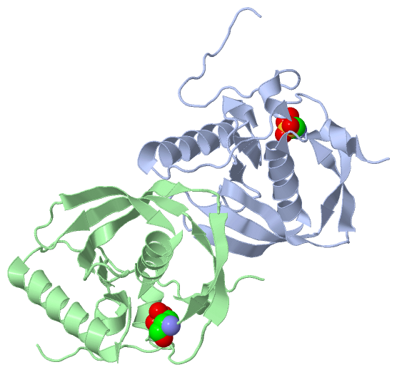
Asymmetric Unit (1, 2)
|
Sites (0, 0)| (no "Site" information available for 1LME) |
SS Bonds (0, 0)| (no "SS Bond" information available for 1LME) |
Cis Peptide Bonds (0, 0)| (no "Cis Peptide Bond" information available for 1LME) |
SAPs(SNPs)/Variants (0, 0)| (no "SAP(SNP)/Variant" information available for 1LME) |
PROSITE Motifs (0, 0)| (no "PROSITE Motif" information available for 1LME) |
Exons (0, 0)| (no "Exon" information available for 1LME) |
Sequences/Alignments
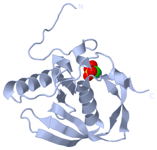
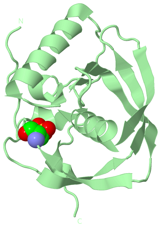
Asymmetric UnitChain A from PDB Type:PROTEIN Length:154 aligned with DEF_THEMA | P96113 from UniProtKB/Swiss-Prot Length:164 Alignment length:154 1 1 11 21 31 41 51 61 71 81 91 101 111 121 131 141 DEF_THEMA - ---------MYRIRVFGDPVLRKRAKPVTKFDENLKKTIERMIETMYHYDGVGLAAPQVGISQRFFVMDVGNGPVAVINPEILEIDPETEVAEEGCLSFPEIFVEIERSKRIKVKYQNTRGEYVEEELEGYAARVFQHEFDHLNGVLIIDRISP 145 SCOP domains d1lmea_ A: Peptide deformylase SCOP domains CATH domains 1lmeA00 A:-8-145 Peptide Deformylase CATH domains Pfam domains ---------------------------------------------------------------------------------------------------------------------------------------------------------- Pfam domains SAPs(SNPs) ---------------------------------------------------------------------------------------------------------------------------------------------------------- SAPs(SNPs) PROSITE ---------------------------------------------------------------------------------------------------------------------------------------------------------- PROSITE Transcript ---------------------------------------------------------------------------------------------------------------------------------------------------------- Transcript 1lme A -8 DKIHHHHHHMYRIRVFGDPVLRKRAKPVTKFDENLKKTIERMIETMYHYDGVGLAAPQVGISQRFFVMDVGNGPVAVINPEILEIDPETEVAEEGcLSFPEIFVEIERSKRIKVKYQNTRGEYVEEELEGYAARVFQHEFDHLNGVLIIDRISP 145 1 11 21 31 41 51 61 71 81 | 91 101 111 121 131 141 87-OCS Chain B from PDB Type:PROTEIN Length:147 aligned with DEF_THEMA | P96113 from UniProtKB/Swiss-Prot Length:164 Alignment length:147 1 | 8 18 28 38 48 58 68 78 88 98 108 118 128 138 DEF_THEMA - --MYRIRVFGDPVLRKRAKPVTKFDENLKKTIERMIETMYHYDGVGLAAPQVGISQRFFVMDVGNGPVAVINPEILEIDPETEVAEEGCLSFPEIFVEIERSKRIKVKYQNTRGEYVEEELEGYAARVFQHEFDHLNGVLIIDRISP 145 SCOP domains d1lmeb_ B: Peptide deformylase SCOP domains CATH domains 1lmeB00 B:-1-145 Peptide Deformylase CATH domains Pfam domains (1) --Pep_deformylase-1lmeB01 B:1-145 Pfam domains (1) Pfam domains (2) --Pep_deformylase-1lmeB02 B:1-145 Pfam domains (2) SAPs(SNPs) --------------------------------------------------------------------------------------------------------------------------------------------------- SAPs(SNPs) PROSITE --------------------------------------------------------------------------------------------------------------------------------------------------- PROSITE Transcript --------------------------------------------------------------------------------------------------------------------------------------------------- Transcript 1lme B -1 HHMYRIRVFGDPVLRKRAKPVTKFDENLKKTIERMIETMYHYDGVGLAAPQVGISQRFFVMDVGNGPVAVINPEILEIDPETEVAEEGcLSFPEIFVEIERSKRIKVKYQNTRGEYVEEELEGYAARVFQHEFDHLNGVLIIDRISP 145 8 18 28 38 48 58 68 78 88 98 108 118 128 138 87-OCS
|
||||||||||||||||||||
SCOP Domains (1, 2)
Asymmetric Unit
 |
CATH Domains (1, 2)
Asymmetric Unit
|
Pfam Domains (1, 2)
Asymmetric Unit
|
Gene Ontology (7, 7)|
Asymmetric Unit(hide GO term definitions) Chain A,B (DEF_THEMA | P96113)
|
||||||||||||||||||||||||||||||||||||||||||||||||||||||
Interactive Views
|
||||||||||||||||||||||||||||||||||||||||||||||||||||||||||||||||||||||||||||||||||||||||||||||||||||||||||||||||||||||||||||||||||||||||||||
Still Images
|
||||||||||||||||
Databases
|
||||||||||||||||||||||||||||||||||||||||||||||||||||||||||||||||||||||||||||||||||||||||||||||||||||||||||||||||||||||||||||||||||||||||||||||||||||||||||||||||
Analysis Tools
|
|||||||||||||||||||||||||||||||||||||||||||||||||||||||||||||
Entries Sharing at Least One Protein Chain (UniProt ID)
Related Entries Specified in the PDB File
|
|