| molecular function |
|---|
| | GO:0030246 | | carbohydrate binding | | Interacting selectively and non-covalently with any carbohydrate, which includes monosaccharides, oligosaccharides and polysaccharides as well as substances derived from monosaccharides by reduction of the carbonyl group (alditols), by oxidation of one or more hydroxy groups to afford the corresponding aldehydes, ketones, or carboxylic acids, or by replacement of one or more hydroxy group(s) by a hydrogen atom. Cyclitols are generally not regarded as carbohydrates. |
| | GO:0008061 | | chitin binding | | Interacting selectively and non-covalently with chitin, a linear polysaccharide consisting of beta-(1->4)-linked N-acetyl-D-glucosamine residues. |
| | GO:0004568 | | chitinase activity | | Catalysis of the hydrolysis of (1->4)-beta linkages of N-acetyl-D-glucosamine (GlcNAc) polymers of chitin and chitodextrins. |
| | GO:0016787 | | hydrolase activity | | Catalysis of the hydrolysis of various bonds, e.g. C-O, C-N, C-C, phosphoric anhydride bonds, etc. Hydrolase is the systematic name for any enzyme of EC class 3. |
| | GO:0016798 | | hydrolase activity, acting on glycosyl bonds | | Catalysis of the hydrolysis of any glycosyl bond. |
| | GO:0046872 | | metal ion binding | | Interacting selectively and non-covalently with any metal ion. |
| | GO:0005515 | | protein binding | | Interacting selectively and non-covalently with any protein or protein complex (a complex of two or more proteins that may include other nonprotein molecules). |
| biological process |
|---|
| | GO:0005975 | | carbohydrate metabolic process | | The chemical reactions and pathways involving carbohydrates, any of a group of organic compounds based of the general formula Cx(H2O)y. Includes the formation of carbohydrate derivatives by the addition of a carbohydrate residue to another molecule. |
| | GO:0016998 | | cell wall macromolecule catabolic process | | The chemical reactions and pathways resulting in the breakdown of macromolecules that form part of a cell wall. |
| | GO:0006032 | | chitin catabolic process | | The chemical reactions and pathways resulting in the breakdown of chitin, a linear polysaccharide consisting of beta-(1->4)-linked N-acetyl-D-glucosamine residues. |
| | GO:0006952 | | defense response | | Reactions, triggered in response to the presence of a foreign body or the occurrence of an injury, which result in restriction of damage to the organism attacked or prevention/recovery from the infection caused by the attack. |
| | GO:0050832 | | defense response to fungus | | Reactions triggered in response to the presence of a fungus that act to protect the cell or organism. |
| | GO:0031640 | | killing of cells of other organism | | Any process in an organism that results in the killing of cells of another organism, including in some cases the death of the other organism. Killing here refers to the induction of death in one cell by another cell, not cell-autonomous death due to internal or other environmental conditions. |
| | GO:0008152 | | metabolic process | | The chemical reactions and pathways, including anabolism and catabolism, by which living organisms transform chemical substances. Metabolic processes typically transform small molecules, but also include macromolecular processes such as DNA repair and replication, and protein synthesis and degradation. |
| | GO:0000272 | | polysaccharide catabolic process | | The chemical reactions and pathways resulting in the breakdown of a polysaccharide, a polymer of many (typically more than 10) monosaccharide residues linked glycosidically. |
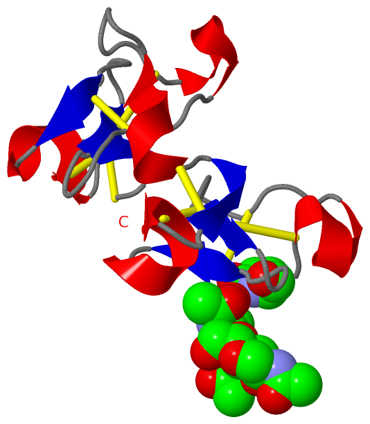
 Description
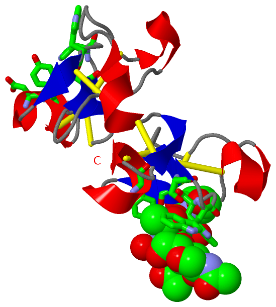